Your selection
Research / 17.12.2025
Observing synapses in action
A team of Berlin-based researchers led by Jana Kroll and Christian Rosenmund has captured the fleeting moment a nerve cell releases its neurotransmitters into the synaptic cleft. Their microscopic images and description of the process are published in “Nature Communications.”
It takes just a few milliseconds: A vesicle, only a few nanometers in size and filled with neurotransmitters, approaches a cell membrane, fuses with it, and releases its chemical messengers into the synaptic cleft – making them available to bind to the next nerve cell. A team led by Professor Christian Rosenmund of Charité – Universitätsmedizin Berlin has captured this critical moment of brain function in microscopic images. They describe their achievement in the journal “Nature Communications.”
Point-shaped connections
“Until now, no one knew the exact steps of how synaptic vesicles fuse with the cell membrane,” says Dr. Jana Kroll, first author of the study and now a researcher in the Structural Biology of Membrane-Associated Processes lab headed by Professor Oliver Daumke at the Max Delbrück Center. “In our experiments with mouse neurons, we were able to show that initially, the process begins with the formation of a point-shaped connection. This tiny stalk then expands into a pore through which neurotransmitters enter the synaptic cleft,” Kroll explains.
“With technology we developed over five years, it was possible for the first time to observe synapses in action without disrupting them,” adds senior author Professor Christian Rosenmund, Deputy Director of the Institute for Neurophysiology at Charité. “Jana Kroll truly did pioneering work here,” says Rosenmund, who is also a board member of the NeuroCure Cluster of Excellence.
The images were produced at the CFcryo-EM (Core Facility for cryo-Electron Microscopy), a joint technology platform operated by Charité, the Max Delbrück Center, and the Leibniz Research Institute for Molecular Pharmacology (FMP) that is directed by Dr. Christoph Diebolder. Also central to the study were Professor Misha Kudryashev, head of the In Situ Structural Biology lab at the Max Delbrück Center, and Dr. Magdalena Schacherl, Project Leader of the Structural Enzymology group at Charité.
Flash-frozen in ethane
To observe synapses in action, the team used mouse neurons genetically modified through optogenetics so they could be activated by a flash of light – prompting them to secrete neurotransmitters immediately. One to two milliseconds after a light pulse, the researchers flash-froze the neurons in liquid ethane at minus 180°C. “All cellular activity stops instantly with this ‘plunge freezing’ method, allowing us to visualize the structures using electron microscopy,” explains Kroll.
The method revealed another intriguing detail: “We found that most of the fusing vesicles were connected by tiny filaments to at least one other vesicle. As soon as one vesicle fuses with the membrane, the next one is already in position,” Kroll reports. “We believe that this direct form of vesicle recruitment enables neurons to send signals over a longer period of time and thus maintain their communication.”
Toward better epilepsy treatment
The vesicle fusion process visualized by the team takes place millions of times a minute in the human brain. Understanding it in detail has important clinical implications. “In many people with epilepsy or other synaptic disorders, mutations have been found in proteins involved in vesicle fusion,” explains Rosenmund. “If we can clarify the precise role of these proteins, it will be easier to develop targeted therapies for these so-called synaptopathies.”
“The time-resolved cryo-electron microscopy approach using light, as we’ve presented here, isn’t limited to neurons,” Kroll adds. “It can be applied across many areas of structural and cell biology.” She now plans to repeat the experiments at the Max Delbrück Center using human neurons derived from stem cells. That won’t be easy, she notes: “In the lab, it takes about five weeks for the cells to develop their first synapses – and they are extremely fragile.”
Text: Anke Brodmerkel
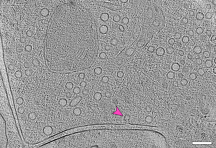
The image captures the moment during which a vesicle (arrow) fuses with the cell membrane. By superimposing several electron microscopy images – a process known as electron tomography – it is possible to see how many vesicles are waiting at the end of a nerve cell to release their biochemical messengers into the synaptic cleft. This space between two nerve cells can be seen as a double line in the image.
© Jana Kroll, Charité / Max Delbrück Center
Source: Joint press release by Charité – Universitätsmedizin Berlin and the Max Delbrück Center
Observing synapses in action
Overview News
News Buch Berlin
Observing synapses in action
A team of Berlin-based researchers led by Jana Kroll and Christian Rosenmund has captured the fleeting moment a nerve cell releases its neurotransmitters into the synaptic cleft. Their microscopic ima...
more ...Eckert & Ziegler Achieves Further Earnings Growth and Double-Digit Sales Growth in the Medical Segment
Eckert & Ziegler SE (ISIN DE0005659700, TecDAX) increased sales in the first nine months of 2025 by 4% to €224.1 million compared to the same period last year. EBIT before special items from continuin...
more ...T-knife Therapeutics Presents Preclinical Data on PRAME-Targeted TK-6302 Highlighting its Potential as a Promising, Category-leading Therapy
Comprehensive TK-6302 data demonstrate preclinical efficacy and safety, supporting clinical readiness, alongside established scalable manufacturing. TK-6302 Clinical Trial Application planned in Q4 20...
more ...Events Buch Berlin
06.01.2026, 16:00
Vorlesung und Lehrerfortbildung: "Wenn das Immunsystem das Gehirn angreift – Autoimmunerkrankungen des zentralen Nervensystems"
Vorlesungsreihe: Neue Wege in der Biomedizin: Aktuelle Forschungsthemen vom Campus Berlin-Buch. Für Lehrkräfte, Schülerinnen und Schüler sowie Interessierte.
more ...20.03.2026, 08:45
Einladung: UniStem Day für Berliner Schülerinnen und Schüler aus Biokursen
Das German Stem Cell Network (GSCN) lädt die an Biologie interessierten Berliner Schülerinnen und Schüler (Biologie-Leistungskurse und Grundkurse) mit ihren Lehrkräften herzlich zum UniStem Day – zum ...
more ...