News
Innovation / 21.07.2026
OMEICOS’ OMT-28 Headed for Phase 3 in Primary Mitochondrial Diseases (PMD) Following Positive End-of-Phase 2 Meeting with the FDA
Berlin, Germany, July 21, 2026
-
Feedback from the U.S. Food and Drug Administration (FDA) supports acceleration of OMT-28’s development path directly into a pivotal Phase 3 study
-
With the vast majority of PMD patients having no FDA-approved therapeutic option, OMT-28 addresses a significant unmet medical need in mitochondrial disease space
-
Results from Phase 2a PMD-OPTION Study were recently presented at Euromit 2026 and
Mito MED 2026, the two largest international conferences on mitochondrial pathologies
OMEICOS, a late-stage clinical biopharmaceutical company developing first-in-class small-molecule therapeutics for mitochondrial and inflammatory disorders, today announced the successful completion of the End-of-Phase 2 (EOP2) meeting with the FDA regarding the development of OMT-28 in Primary Mitochondrial Diseases (PMD). During the meeting, the FDA expressed strong support for OMEICOS’ plans to advance the program into
late-stage clinical development and provided clear, constructive guidance to structure the next steps efficiently. As a result, OMEICOS is now positioned to advance its proprietary, fully owned lead product candidate OMT-28 directly into a pivotal Phase 3 study.
The EOP2 meeting with the FDA was supported by results from the successfully concluded multicentre, open-label Phase 2a PMD-OPTION Study in patients with PMD. The PMD-OPTION Study demonstrated
OMT-28’s therapeutic potential to improve physical condition in patients with PMD based on significant recovery of the impaired mitochondrial function in the responding patients. The study also further underscored the excellent safety and tolerability profile of OMT-28, which has been evaluated in more than 190 individuals to date.
OMT-28 has the potential to be a first-in-class and best-in-class therapy and is clearly differentiated from competing approaches in PMD through its unique dual mechanism, targeting both redox balance and mitochondrial restoration, addressing core disease biology more comprehensively than single-pathway approaches. OMT-28 leverages the activation of mitochondrial sirtuin family members SIRT1 and SIRT3, mainly through biased modulation of S1PR1 (Sphingosine-1-Phosphate Receptor 1) signalling.
As a once-daily oral small molecule, OMT-28 would offer superior convenience and adherence compared to currently evaluated injectables or twice-daily future alternatives. Moreover, cardiomyopathy patients—a subgroup often excluded in other clinical development programs—highlights OMT-28’s potentially broader target population further reinforcing its best-in-class product profile. Beyond PMD, OMEICOS is pursuing a ‘pipeline-in-a-drug’ strategy with OMT-28 and sees potential for expansion into larger disease areas.
“The strong endorsement from the FDA marks a significant milestone for OMEICOS as a company and for our mission to address Primary Mitochondrial Disease. The meeting’s outcome even surpassed our expectations, as we are now able to advance directly from our concluded Phase 2a study into a pivotal Phase 3 study,” said Dr. Robert Fischer, CEO/CSO of OMEICOS Therapeutics. “Our goal now is to bring a much-needed new treatment option into the final stages of development—and hopefully into medical practice—as quickly as possible. We are currently evaluating the best possible infrastructure, strategic partners, and resources to achieve this goal efficiently.”
PMD patients suffer from debilitating and life-threatening health consequences, such as severely limited physical stamina and disease-related changes in the heart and skeletal muscles, as well as associated neurological disorders. The disease represents a heterogeneous group of conditions including the most prevalent subtypes MELAS, non-MELAS, and MIDD. The upcoming pivotal Phase 3 study is planned to enrol up to 160 adult patients stratified by these three common PMD subgroups and will evaluate the efficacy of OMT-28 at a 24 mg once-daily dose over 24 weeks, with an option to extend treatment up to 104 weeks. The trial will employ an adaptive design, allowing adjustments to sample size and treatment duration based on interim analyses, ensuring flexibility and efficiency in this rare disease setting. The primary endpoint is a composite measure combining the 12-Minute Walk Test (12MWT) and the 5x Sit-to-Stand Test (5xSST), with secondary endpoints including quality-of-life assessments and exploratory biomarkers such as NAD⁺, GSH, and their ratios. This design reflects the FDA’s constructive guidance and underscores the potential of OMT-28 to transform the treatment landscape for patients in need.
About OMEICOS
OMEICOS Therapeutics has discovered a series of metabolically robust synthetic analogues of omega-3 fatty acid-derived epoxyeicosanoids that have the potential to treat mitochondrial dysfunction, inflammatory, cardiovascular and other diseases. Epoxyeicosanoids activate cell type-specific endogenous pathways that promote organ and tissue protection. OMEICOS’ small molecules are orally available and show improved biological activity and pharmacokinetic properties compared to their natural counterparts. The Company’s most advanced development program OMT-28 is headed for a pivotal Phase 3 clinical study in Primary Mitochondrial Diseases (PMD). For more, please visit: www.omeicos.com
Contacts
OMEICOS Therapeutics GmbH
Dr. Robert Fischer, CEO, CSO
Phone: +49 (0) 30 9489 4810
E-Mail: r.fischer@omeicos.com
www.omeicos.com
Quelle: Omeicos Therapeutics GmbH
Research / 20.07.2026
A single cocaine dose alters mouse brain cells
Researchers led by Ana Pombo have found that just one exposure to the drug can create changes in mouse brain cells that persist for at least two weeks. Their findings were presented at the Federation of European Neuroscience Societies Forum on July 7, 2026.
Cocaine is a highly addictive drug that can cause anxiety and paranoia in users, and can lead to heart damage, impotence and poor mental health in the long term. According to the U.N Office on Drugs and Crime, cocaine use is at an all-time high, with an estimated 25 million users worldwide.
“We know that cocaine hijacks the reward machinery of the brain,” said Dr. Ana Pombo, Bloomberg Distinguished Professor at Johns Hopkins University, during the Federation of European Neuroscience Societies (FENS) Forum on July 7, 2026. Pombo also has a joint appointment as Guest Group Leader of the Epigenetic Regulation and Chromatin Architecture lab at the Berlin Institute of Medical Systems Biology of the Max Delbrück Center. “Most people do not become addicted after using cocaine once, but many do after a second use or repeated exposures. However, we don’t know enough about what is happening to brain cells exposed to cocaine and whether these effects are long-lasting.”
Her team has been using mice to see where the brain stores the memory of taking cocaine for the first time, and to understand why addiction occurs after repeated use, even when cocaine use is months or years apart.
Mapping effects in the brain
The researchers used a technique called genome architecture mapping to understand the effects of cocaine on mouse brains. This approach makes it possible to study how genetic material is organized inside a cell. Although genes provide a blueprint for all cells in the body, their three-dimensional organization can dictate when genes are switched on or off in any individual cell.
Compared to mice not exposed to cocaine, researchers found that the 3D structure of the genome was extensively altered in brain cells called dopaminergic neurons in the ventral tegmental region of the midbrain. This part of the brain is known to play an important role in reward and motivation. The changes could be seen 24 hours after exposure to cocaine, and persisted. Some changes were even greater two weeks after exposure.
For example, among these changes, they found that a single cocaine exposure prompts the development of around 1,700 new ‘chromatin domain insulation areas’ — parts of the genome that can help regulate the activity of genes — and the loss of around another 1,100 of these areas.
The researchers also looked in detail at which genes were active and which were inactive in mouse brain cells exposed to cocaine compared with brain cells not exposed to cocaine.
This showed that exposed cells were producing more of some of the brain’s signalling molecules, called neuropeptides, that have been linked to addiction in humans. Other genes that help the brain cell function normally had become less active.
Implications for addiction
“Our results suggest that a single exposure to cocaine ‘rewires’ the genome of these important brain cells,” says Pombo. “The fact that we found such big changes that persist for two weeks is unexpected and it suggests that the drug is leaving a longer-term ‘scar’ in the genome of the brain cells.”
She adds: “These persistent changes may be setting the stage for a stronger response after a second dose of cocaine, which could help explain why the brain becomes susceptible to cocaine addiction. We still need to investigate how long these changes last. Are they permanent, or can the brain cells recover over time? We also need to investigate how these changes translate to the risk of addiction.”
“Cocaine use is a serious and growing problem around the world. We need to understand the effects of this drug and how people become addicted, but it’s almost impossible to study these mechanisms in detail in the human brain, so instead we look at mice,” said Professor Christina Dalla from the National and Kapodistrian University of Athens, Greece, chair of the FENS Forum communication committee and who was not involved in the research.
“In this study, scientists have identified profound and lasting changes in mouse brain cells after just one exposure to cocaine,” she added. “This shows that cocaine can alter the structure of the genome in these cells and this alteration may persist over time. These findings challenge the idea that occasional recreational use of cocaine may be harmless as they suggest that one use could change our brains and raise the risk of addiction in the future.”
Researching these changes in greater detail could help us understand why some people are more likely than others to become addicted. This could also help us to find new ways to treat addiction.
Text: FENS Forum 2026
https://www.mdc-berlin.de/news/Research / 17.07.2026
How the skin distinguishes cool from warm
Researchers in the lab of James Poulet have uncovered how the nervous system senses cool and warm temperatures. The findings, published in “Neuron” challenge a long-standing view of temperature sensing and could guide future research into pain and sensory disorders.
Whether we hold a warm mug or step onto a cool floor, specialized nerve cells in the skin constantly report temperature to the brain. Scientists have long assumed that separate groups of sensory cells detect non-painful cool and warm temperatures. Now researchers led by Drs. Phillip Bokiniec and Clarissa Whitmire in the Neural Circuits and Behavior Lab of Dr. James Poulet at the Max Delbrück Center have found that this assumption is too simplistic.
“Rather than relying on separate “warm” and “cool” sensors, we found that the nervous system appears to use one population of cells that signals both directions of temperature change,” explains Bokiniec, who shares first authorship of the study with Whitmire. Bokiniec is a now researcher in the Sensory Neural Coding lab of Dr. Clarissa Whitmire at the Queensland Brain Institute.
Using advanced imaging in mice, the team report in “Neuron” that most temperature-sensitive nerve cells are activated by cooling, and merely reduce their activity when the skin warms. The researchers also showed that these cells react to the actual temperature of the skin rather than simply detecting how much it has changed. This finding reshapes scientists’ understanding of one of the body’s most fundamental senses.
“Scientists have known about these neurons for years,” notes Poulet, “but they were thought to be relatively rare. What surprised us was discovering that they make up most of the temperature-sensing cells.”
Imaging neurons in live mice
The researchers developed a method to image hundreds of temperature-sensing nerve cells in the spinal sensory ganglia of awake mice over time. They gently warmed and cooled the animals’ paws while recording the activity of individual neurons using two-photon microscopy. They also performed this experiment in anesthetized mice and found the same result. This proved that that the anesthetic itself did not affect their results.
The team then selectively blocked or activated temperature-sensitive ion channels. Blocking the protein TRPM8 — long known as the body’s main sensor for detecting cool temperatures — eliminated both the response to cooling and the dampening effect that warming has on these nerve cells. This showed that a single molecular sensor can generate signals for both cool and warm, challenging the traditional view that separate receptors are needed for each sensation.
The team also developed a computer model to test their hypothesis. It showed that simply changing the activity of a TRPM8 was enough to reproduce the different response patterns seen in the experiments.
Understanding sensory disorders
Temperature sensation is essential for everyday life, but it is also disrupted in many medical conditions, including neuropathic pain, diabetic neuropathy, chemotherapy-induced nerve damage and disorders that cause abnormal sensitivity to cold. “Understanding how healthy temperature sensing works is a prerequisite for understanding what goes wrong in disease,” says Whitmire.
The researchers next plan to investigate how these signals are processed in the spinal cord, how painful temperatures are encoded, and whether the same principles apply in humans.
Text: Gunjan Sinha
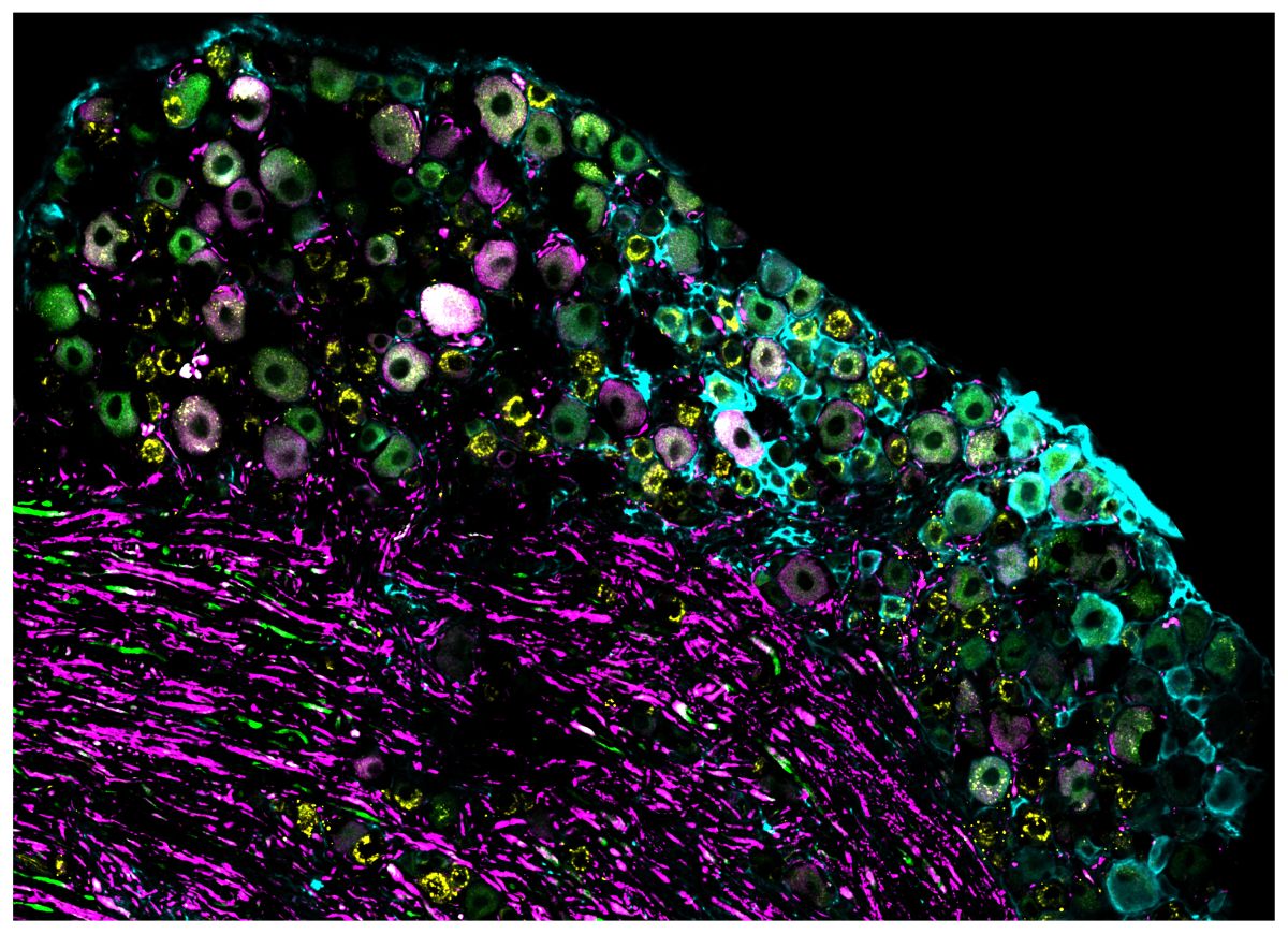
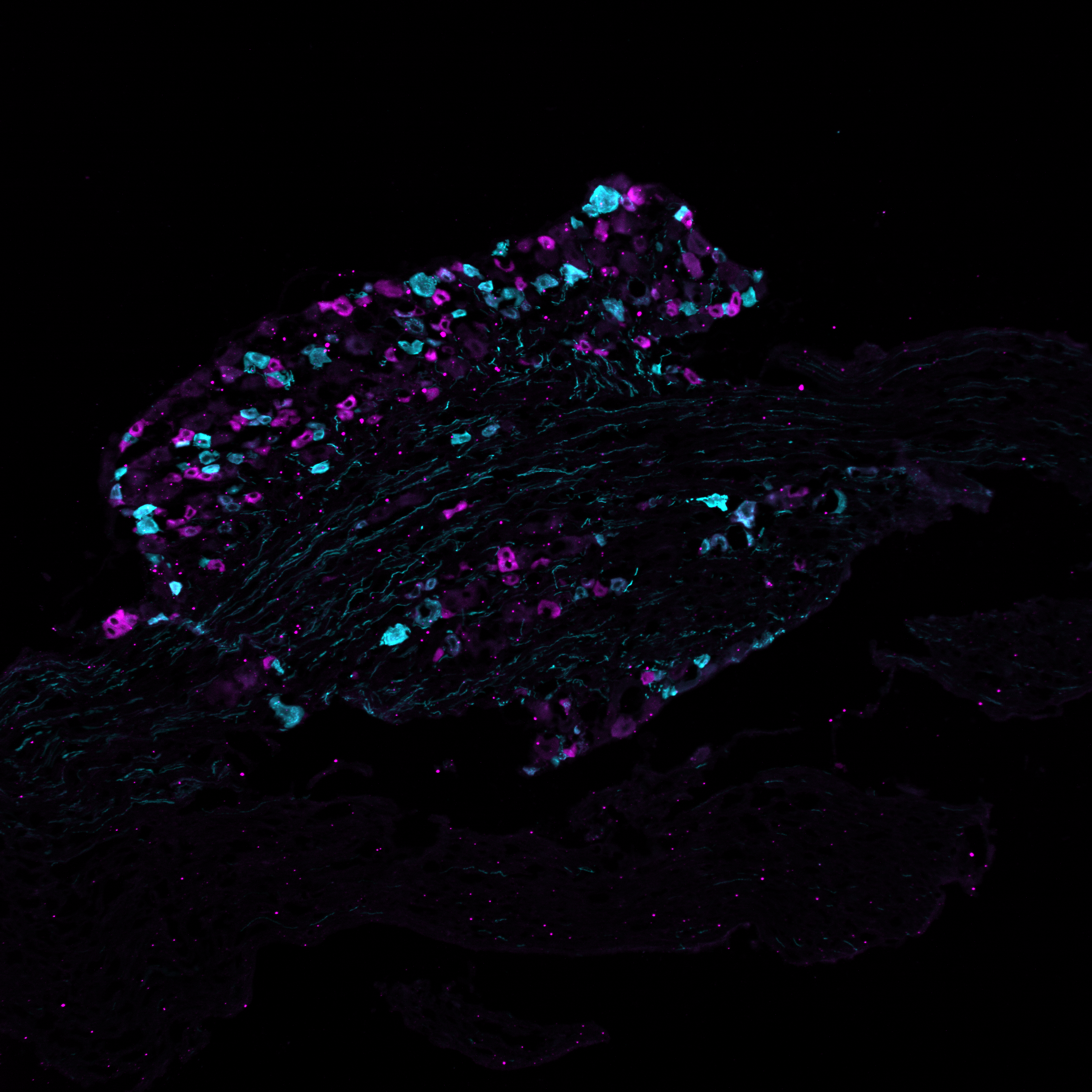
Figure: A cluster of nerve cells from a mouse that carries sensory information from the hind leg to the spinal cord. Some of these nerve cells detect temperature, while others respond to different sensations such as touch or pain. The different colors highlight specific groups of cells.
Further information
Research, Innovation, Patient care / 07.07.2026
A Leap into the World of Big Pharma
The Successful Transformation of a Startup: An Interview with Dr. Christian Regenbrecht, Vice President of Translational Oncology at GSK and Founder of CELLphenomics
Your startup, CELLphenomics, has been successfully conducting pharmacological research on patient-derived tumor organoids at the Berlin-Buch campus for more than ten years. It has been part of GlaxoSmithKline (GSK) since 2025. What was the background to this?
3D cell culture models, also known as organoids, are becoming increasingly sophisticated and reliable for testing therapeutic compounds. In certain cases and when supported by robust data, regulatory authorities are even willing to forgo animal testing. These models play a key role in making new, better drugs available more quickly. Our startup has advanced organoid research and earned an international reputation. We were the first commercial biobank in Germany to receive ISO certification. We cleared this hurdle with such flying colors that we have since been helping to shape, as part of the ISO commission, how pharmaceutical research with organoids will be conducted in the future.
We had already carried out joint projects with GSK. They were impressed by our research, the high quality of our data, and the volume of data and models we produced. The rule is: The higher the data quality, the higher the predictive value, and the better the foundation for machine learning. They wanted to bring that expertise into their company.
What motivated you to sell?
GSK’s corporate culture was a good fit for us, and ultimately, the deciding factor for me was that together we could have a far greater impact. We share their commitment to putting patients first and providing them with the best possible medications as quickly as possible.
Now we not only have greater resources at our disposal, but we’re also getting much more of a hearing from regulatory agencies like the FDA and EMA. Another very important point: Our entire team remained intact under the new structure. All 18 of us have good, secure jobs.
How did the transition to becoming part of GSK go?
It wasn’t easy at all to preserve the core and identity of CELLphenomics—that is, the reason why people enjoy coming to work here—while still adopting GSK’s corporate culture. It involved many small details, such as strict occupational safety requirements or the shorter work hours typical in England. Overall, there’s a much greater need for coordination in an international pharmaceutical company. They look very closely at whether things are actually needed to address the company’s most pressing issues.
Many things are becoming easier within GSK’s organizational structure: Thanks to legally sound contracts with international partners, for example, we can use tissue from all over the world to grow organoids for our projects.
We’ve expanded, upgraded our labs, and acquired a lot of new laboratory equipment. This allows us to conduct even better research, offer higher throughput, and deliver even better quality. GSK’s strong structure enables continuity—we’re working at full capacity, but no longer partially overburdened as we were in the startup phase.
Has your research changed?
Yes, I would definitely say so. GSK has many collaborations and partnerships. We are now part of a transnational tumor network in which leading scientists in Tokyo, Oxford, and Cambridge discuss joint projects and research questions, evaluate data, and design new experiments. We benefit enormously from this.
How are you settling into your new role?
I have time again to read papers, think about the best possible solution to a scientific question, and develop exciting projects.
Because GSK’s strong support team backs us up in every way, I don’t have to worry as much about things like tax returns, legal issues, or getting the best offers from manufacturers. I was even able to take three weeks of vacation.
ASC Oncology was your second startup, which offered personalized drug testing on tumor organoids.
We no longer help individual patients, but rather a group of individuals. As part of clinical development, we can contribute our expertise in a much more structured way and ensure that active ingredients reach the market faster.
Looking back at your early days?
In 2014, we were a very small startup in a “Shared Lab.” How unlikely was the transformation into something much bigger? Many startups fail during this early growth phase in today’s increasingly complex, globalized world—and no one is to blame for that.
We’ve always been very well supported—even through the straightforward, quick solutions provided by campus management.
Text and Photo: Christine Minkewitz / Campus Berlin-Buch GmbH
Dieses Interview erschien zuerst im Standortjournal buchinside 02/2026.
Innovation / 24.06.2026
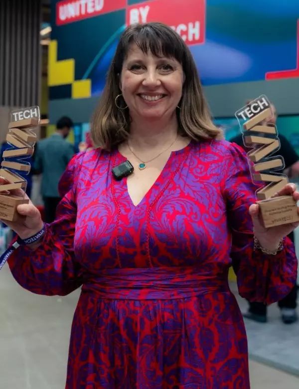
Alithea Bio Scores Double Victory at VivaTech 2026 for Breakthrough in Preclinical Safety
PARIS / BERLIN / FREIBURG — 23.6.2026
Deep-tech biotech innovator Alithea Bio has achieved a spectacular double victory at VivaTech 2026, Europe’s largest startup and tech event, taking home both the coveted “Tech for Change” Jury Prize and the Audience Favorite Award. Selected by an elite panel of global investors and corporate leaders—and backed by overwhelming public support—the awards recognize Alithea Bio’s pioneering work in eliminating toxicity risks at the preclinical stage to dramatically increase clinical trial success rates.
Historically, promising cancer therapies and vaccines fail during human trials due to unforeseen toxicities and adverse immune reactions that standard models miss. Alithea Bio’s proprietary platform, HLA-Compass AI, solves this multi-billion-dollar bottleneck during preclinical development by mapping complex Human Leukocyte Antigen (HLA) interactions. Unlike purely digital AI startups, Alithea Bio utilizes a unique hybrid model—combining its own active wet lab with an advanced computational engine to accurately predict and filter out toxicity risks long before an asset ever enters human clinical trials.
The company’s commercial traction matches its scientific breakthrough. Fully bootstrapped to date, Alithea Bio amassed 22 million proprietary datapoints and generated €1.6M in revenue in 2025 through active preclinical validation partnerships with global pharmaceutical leaders.
“Sweeping both the jury and audience awards at VivaTech is an incredible validation of our mission, our impact and our scalable commercial model. Too many life-saving therapies are derailed late in development because traditional preclinical models fail to predict human toxicity. HLA-Compass isn’t just about saving pharma companies millions in failed trials; it’s about ensuring that only the safest, most precise assets advance to human clinical trials. The crowd and the jury both sent a clear message: the industry is ready for this change,” says Fanny Giannou, CEO of Alithea Bio.
The double accolade comes at a pivotal moment of global scaling for Alithea Bio. The company is currently expanding its strategic partnerships with global pharmaceutical leaders and launching an institutional Seed funding round to transition its proven data-service capabilities into a highly scalable software platform while expanding its footprint into the US market.
For more information about Alithea Bio and the HLA-Compass platform, visit www.alithea-bio.com or contact news@alithea-bio.com.
About Alithea Bio
Alithea Bio is a Freiburg/Berlin-based biotechnology company dedicated to transforming the safety and predictability landscape of preclinical oncology and vaccine therapeutics. By utilizing advanced predictive modeling driven by a 22-million node wet-lab and AI hybrid data engine, Alithea Bio enables pharmaceutical developers to identify and mitigate toxicity risks at the preclinical stage, ensuring safer, more effective treatments successfully advance through clinical pipelines to reach patients worldwide.
Media Contact
Fanny Giannou
CEO, Alithea Bio
news@alithea-bio.com
Innovation / 24.06.2026
Eckert & Ziegler SE Annual General Meeting Approves Significantly Higher Dividend
The Annual General Meeting of Eckert & Ziegler SE (ISIN DE0005659700) today approved the proposal of the Executive Board and Supervisory Board and resolved to pay a dividend of € 0.22 per share (previous year: € 0.17) for the 2025 fiscal year. As in previous years, the Annual General Meeting was held as an in-person event, right next to the Eckert & Ziegler SE headquarters in Berlin. In total, 55.37 % of the company’s share capital was represented. The Annual General Meeting endorsed the members of the Executive Board and the Supervisory Board of Eckert & Ziegler SE for the 2025 fiscal year and approved all items on the agenda by a large majority.
The detailed voting results of the Annual General Meeting and the CEO’s presentation are available on the Eckert & Ziegler SE website:
https://www.ezag.com/investors/annual-general-meeting/
About Eckert & Ziegler
Eckert & Ziegler SE with more than 1.000 employees is a leading specialist for isotope-related components in nuclear medicine and radiation therapy. The company offers a broad range of services and products for the radiopharmaceutical industry, from early development work to contract manufacturing and distribution. Eckert & Ziegler shares (ISIN DE0005659700) are listed in the TecDAX index of Deutsche Börse.
Contributing to saving lives.
Research / 23.06.2026
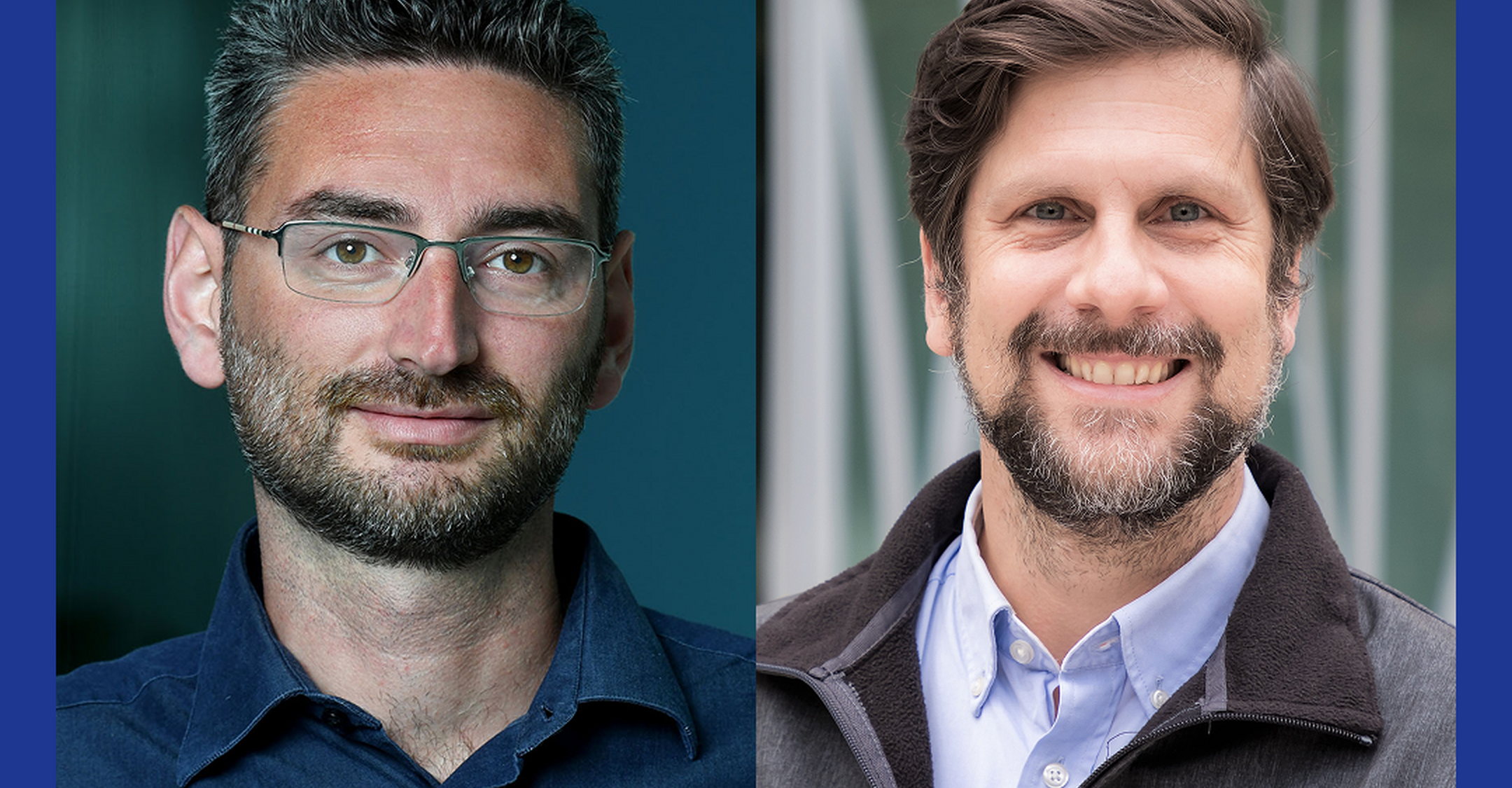
ERC Advanced Grants awarded to two Berlin researchers
ERC Advanced Grants awarded to two Berlin researchers
Uwe Ohler and Gaetano Gargiulo have been awarded prestigious ERC Advanced Grants. With funding of €2.5 million each, they will spend the next five years investigating how protein synthesis is regulated and identifying vulnerabilities in pediatric brain tumors.
Two Max Delbrück Center scientists have secured prestigious European Research Council (ERC) Advanced Grants, one of Europe’s highest honors for established researchers: Dr. Uwe Ohler, Group Leader of the Computational Regulatory Genomics lab, and Dr. Gaetano Gargiulo, Group Leader of the Molecular Oncology lab.
The ERC supports ambitious projects by outstanding researchers who have already achieved significant success in their fields. Recipients receive up to €2.5 million over five years to pursue promising research ideas. This year only 319 researchers were selected from 3,329 who applied from across Europe.
Designing RNA with precision
Ohler won funding for project TRANS-DECODE. With his team, he will investigate how cells regulate translation — the process by which cells use the messenger RNA transcripts of our genetic material — to produce proteins. “Many genetic diseases are not caused by changes in protein-coding genes but by errors in translation,” says Ohler. “We want to better understand this finely tuned process in which regulatory regions in messenger RNA play an important role, and develop molecular tools that allow us to intervene in a more targeted way.”
The Ohler lab will combine machine learning — a type of artificial intelligence (AI) — with advanced molecular biology techniques. These methods enable researchers to measure the activity of thousands of regulatory RNA segments simultaneously, modify them in very specific ways, and generate snapshots of all proteins produced in a cell.
The team will use explainable AI (XAI) to find regulatory elements hidden within messenger RNA and to understand how manipulating them affects translation. “With XAI, we can ensure that the models’ predictions remain transparent and understandable to us,” explains Ohler. For their experiments, the researchers will use both human cells and zebrafish, which serve as a vertebrate model.
“With TRANS-DECODE, we not only want to unravel the molecular logic of translation step by step, we also want to identify new ways to correct defects in regulatory RNA segments that contribute to human disease,” says Ohler. “Our long-term goal is to rationally design RNA molecules for both therapeutic and synthetic biology applications, such as developing better vaccines.”
Avatars of brain tumors
Gargiulo will use his grant to create highly realistic models of pediatric brain tumors in a project called MOIRA. He and his team plan to replicate in detail the transformation of a healthy, maturing brain cell into a tumor cell within a brain organoid — a type of miniature organ. These tumor avatars will help his team better understand how these cancers develop and identify potential treatment strategies.
The grant marks Gargiulo’s fourth ERC award. He won a Starting Grant in 2016, which he used to begin creating models of brain tumors. This was followed by two Proof of Concept grants in 2022 and 2024 that helped his team further develop a new technology called synthetic genetic tracing. The method uses artificial DNA molecules, called reporter genes, to visualize specific cellular activities within tissues.
In MOIRA, these two fields of research are now being brought together: Gargiulo and his colleagues aim to develop reporter genes that turn on as soon as a cell activates a tumor-like program. “In human brain organoids, the reporter genes will help us determine which cells exhibit characteristics of brain tumors, which type of brain tumor they resemble, and when these changes occur,” explains Gargiulo. His team plans to purify the cells and use them to develop models that, step by step, increasingly resemble a real tumor. The project will combine synthetic biology with organoid research and AI-supported validation.
“With MOIRA, we want to reconstruct exactly how pediatric brain tumors arise,” says Gargiulo. “If we succeed in creating faithful tumor avatars, we can say that we have truly come much closer to understanding these diseases. In addition, our models will allow us to systematically search for tumor vulnerabilities — and thus identify potential targets for new drugs.”
Text: Anke Brodmerkel
Further information
Computational Regulatory Genomics
Photo left side: Gaetano Gargiulo © David Ausserhofer, Max Delbrück Center
Photo right side: © Felix Petermann, Max Delbrück Center
Innovation / 11.06.2026
Eckert & Ziegler and DC Pharma Open Commercial Medical Isotope Production Site in Jintan
Eckert & Ziegler SE (ISIN DE0005659700, TecDAX) today officially opened the new medical isotope production site of its joint venture Qi Kang Medical Technology (Changzhou) Co., Ltd. (QKM) in the Jintan district of Changzhou, China. With the opening, Eckert & Ziegler and its Chinese partner DongCheng Pharma (DC Pharma) take a decisive step toward local supply of medical isotopes for cancer diagnostics and therapy in the growing Chinese market. Production will start with Germanium-68 (Ge-68), the parent isotope used to produce Gallium-68 (Ga-68), a crucial isotope for diagnostic imaging.
Around 60 invited guests attended the opening ceremony, including representatives of both joint venture partners, as well as government officials. To support production, the company acquired and successfully installed a cyclotron in late 2025. The facility offers 9,500 m² of usable floor space, and production is scheduled to begin in early 2027. In a planned second phase, the site will also become the first in China dedicated to the commercial production of Actinium-225 (Ac-225), a key isotope for next-generation targeted cancer therapies.
"The opening of our site in Jintan is an important milestone for our growth strategy in China and for Eckert & Ziegler's position as a global supplier of vital radioisotopes," said Dr. Harald Hasselmann, CEO of Eckert & Ziegler SE. "Producing Ge-68 locally gives clinicians across China a reliable domestic supply of the parent isotope behind the Ga-68 generators that power modern PET diagnostics. This reflects the kind of integrated, end-to-end capability we have built over decades."
“For China, the Jintan site marks a turning point," said Zhigang Luo, Group CEO of DC Pharma. "With local production of Ge-68 and, in the next phase, Ac-225, we are bringing a steady supply of key medical isotopes to Chinese patients and strengthening the resilience of the entire nuclear medicine value chain. Together with Eckert & Ziegler, we are laying the foundation for a new generation of precision diagnostics and therapies in China."
Eckert & Ziegler reliably supplies Gallium-68, Lutetium-177, Yttrium-90, and Actinium-225 to leading pharmaceutical companies and research institutions worldwide. With expertise in radioisotope production as well as global logistics and CDMO services, the company is committed to continuously supporting the development and delivery of innovative radiopharmaceuticals.
About Qi Kang Medical Technology (Changzhou) Co., Ltd. (QKM)
Qi Kang Medical Technology (Changzhou) Co., Ltd. (QKM) is a 50:50 joint venture between Eckert & Ziegler SE and DongCheng Pharma headquartered in the Jintan district of Changzhou, China. QKM operates a state-of-the-art production site for cyclotron-based medical isotopes including Germanium-68 and is committed to establishing the first commercial Actinium-225 production in China. The venture is dedicated to supplying the Chinese radiopharmaceutical market with key radioisotopes for cancer diagnostics and therapy.
About Eckert & Ziegler
Eckert & Ziegler SE, with more than 1,000 employees, is a leading specialist in isotope-related components for nuclear medicine and radiation therapy. The company offers a broad range of services and products for the radiopharmaceutical industry, from early development work to contract manufacturing and distribution. Eckert & Ziegler shares (ISIN DE0005659700) are listed in the TecDAX index of Deutsche Börse.
Contributing to saving lives.
About DongCheng Pharma
Yantai Dongcheng Pharmaceutical Group Co., Ltd. (DC Pharma), founded in 1998 and headquartered in Yantai, China, is one of the country's leading pharmaceutical groups and a recognized leader in nuclear medicine in China. The group develops, manufactures and sells biochemical active pharmaceutical ingredients, finished dosage forms, nuclide drugs and health products for therapeutic areas including oncology, cardiovascular, urology and orthopedics, and exports its products to more than 40 countries. DC Pharma is listed on the Shenzhen Stock Exchange (002675.SZ).
Source: Pressemitteilung Eckert & Ziegler SE
Eckert & Ziegler and DC Pharma Open Commercial Medical Isotope Production Site in Jintan
Research, Innovation, Patient care / 22.05.2026
Berlin honors outstanding deep-tech companies—including MyoPax
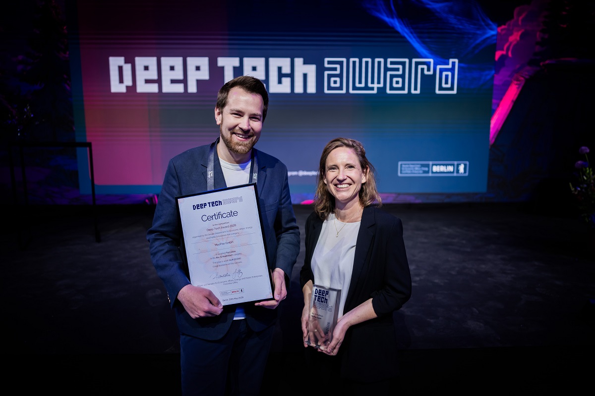
This year, for the first time, a Deep Tech Award was presented in the Bio & Health Tech category. The winner is MyoPax, a spin-off of the Max Delbrück Center and Charité–Universitätsmedizin Berlin.
AI-powered fact-checking to combat disinformation, novel cell therapies for muscle tissue regeneration, sustainable 3D printing, and decentralized satellite communications: On the evening of Wednesday, May 20, 2026, the Senate Department for Economic Affairs, Energy and Public Enterprises presented the Deep Tech Award to five outstanding Berlin-based companies. For the eleventh consecutive year, the administration is thus recognizing technological, research-based innovations from Berlin that stand out for their practical application, societal relevance, and added value. The awards ceremony, attended by Senator for Economic Affairs Franziska Giffey and Permanent Secretary for Economic Affairs Michael Biel, took place for the first time as part of the Deep Tech Momentum conference at Wilhelm Studios in Berlin.
Franziska Giffey, Mayor and Senator for Economic Affairs, Energy and Public Enterprises: “We want to make Berlin the number one hub for innovation in Europe. The companies honored today are turning cutting-edge technological research into practical applications and offering solutions to current challenges. In doing so, they impressively demonstrate the immense innovative potential that Berlin possesses. With the Deep Tech Award, we recognize the courage and excellence of our founders. Through our new partnership with the Deep Tech Momentum conference, we are also creating a platform that connects Berlin’s brightest minds even more closely with industry partners and international investors. In doing so, we are ensuring sustainable growth and technological sovereignty for Berlin as a hub.”
The Deep Tech Award offers a total prize pool of 50,000 euros and is presented in five categories: “Advanced Manufacturing,” “Bio- & Healthtech,” “Artificial Intelligence,” “Quantum Technologies, Photonics & Microelectronics,” and “Web3 & Distributed Ledger Technologies (DLT).” From over 84 applications, expert juries selected five winning companies, each of which will receive prize money of 10,000 euros.
With the Deep Tech Award being integrated into Deep Tech Momentum for the first time, the prize is being positioned even more strongly on the international stage. The conference is considered one of Europe’s leading platforms for connecting deep-tech startups with companies and investors. The new partnership underscores the award’s mission to not only honor Berlin’s most innovative technology companies but also to connect them even more closely with the European innovation ecosystem. In this context, the special “Deep Tech Award for Breakthrough Momentum” was also presented for the first time this year. It is aimed at European startups curated by Deep Tech Momentum that combine excellent scientific innovation with exceptional scaling potential.
The winners of the 2026 Deep Tech Award at a glance:
Deep Tech Star in the “Advanced Manufacturing” category: Endless Industries GmbH
Endless Industries is revolutionizing the manufacturing of fiber-reinforced composites—materials in which carbon fibers are embedded in a binding resin—with a 3D printing solution. This makes it possible to replace complex and expensive manufacturing processes and reduce waste. Website:
www.endless.industries
Deep Tech Star in the “Bio- & Healthtech” category: MyoPax GmbH
MyoPax’s work focuses on innovative cell therapies and gene corrections for the regeneration of muscle tissue. In doing so, the company specifically targets severe muscle injuries and diseases, offering patients promising new treatment options. Website
: www.myopax.com
Deep Tech Star in the “Artificial Intelligence” category: Gretchen AI GmbH
Gretchen AI develops state-of-the-art AI to detect deepfakes and fake news and can reconstruct their dissemination history. This enables the Berlin-based company to help major media organizations conduct fact-checks up to six times faster while maintaining the same level of reliability—a crucial contribution to safeguarding the public information space.
Website: www.gretchen-ai.com
Deep Tech Star in the “Quantum Technologies, Photonics & Microelectronics” category: Xavveo GmbH
Xavveo develops photonic radar sensors that set new standards in fields such as navigation and measurement technology, for example in environmental sensing for automobiles. The technology enables unprecedented precision and has the potential to fundamentally replace existing sensor solutions across a wide range of industries.
Website: www.xavveo.com
Deep Tech Star in the “Web3 & Distributed Ledger Technologies (DLT)” category: Decen Space UG
The startup Decen Space is developing a decentralized coordination network consisting of software and hardware components for the secure and efficient synchronization of data streams between satellites and ground stations. This solution enables higher data transfer rates at significantly lower costs and also allows satellite operators more contact time with their satellites. Website
: www.decenspace.com
Deep Tech Star of the Special Prize “Deep Tech Award for Breakthrough Momentum”: Six Robotics AS
Six Robotics AS is a Norwegian company specializing in the development of autonomy software for fleets of unmanned aerial vehicles. The software is based on innovative swarm intelligence algorithms and real-time mission control architectures that ensure a high degree of autonomy and efficiency for the aircraft. This enables the company’s drones to coordinate and execute missions as intelligent teams. As a result, the use of networked autonomous systems in modern defense operations is being advanced. Website: www.sixrobotics.com
For more information about Deep Tech Berlin and the Deep Tech Award, please visit: www.berlin.de/deeptech/
Source: Press release of the Senate Department for Economic Affairs, Energy and Public Enterprises
Innovation / 22.05.2026
Eckert & Ziegler Receives “Best Managed Companies Award” Once Again
Eckert & Ziegler SE (ISIN DE0005659700) has won the “Best Managed Companies Award” for the third time in a row. With this honor, Deloitte Private, UBS, Frankfurter Allgemeine Zeitung, and the Federation of German Industries (BDI) recognize excellently managed medium-sized companies.
“Good corporate leadership is of central importance, especially in economically challenging times. The Best Managed Companies Award is a well‑deserved recognition for companies that successfully combine responsibility, foresight, and sustainable action,” says Tobias Vogel, CEO of UBS Europe SE.
“This award is both recognition and motivation for us. It confirms that we are on the right path with a clear strategy in the growing nuclear medicine market—together with a fantastic team around the world,” added Dr Dirk W. Becker, a member of the Group Executive Committee of Eckert & Ziegler SE, who accepted the corporate trophy on behalf of the company at the awards ceremony in Frankfurt am Main.
The award is the result of a comprehensive, multi-stage application process in which companies are assessed for their excellence in the core areas of strategy, productivity and innovation, culture and commitment as well as finance and governance. A consistently high level of performance in all four categories is a prerequisite for selection. The final decision is made by an independent jury made up of renowned experts from business, science and the media.
About Eckert & Ziegler.
Eckert & Ziegler SE with more than 1,000 employees, is a leading specialist for isotope-related components in nuclear medicine and radiation therapy. The company offers a broad range of services and products for the radiopharmaceutical industry, from early development work to contract manufacturing and distribution. Eckert & Ziegler shares (ISIN DE0005659700) are listed in the TecDAX index of Deutsche Börse.
Contributing to saving lives.
Innovation / 12.05.2026
Eckert & Ziegler with a Successful Start to the Year. 2026 Forecast Confirmed.
1st Quarter 2026:
- Sales of €72.9 million (previous year: €68.2 million)
- EBIT before special items of €16.0 million (previous year: €16.2 million)
- Net income of €10.4 Mio. (previous year: €9.7 million)
Forecast 2026:
- Sales of around €320 million (confirmed)
- EBIT before special items of around €80 million (confirmed)
Eckert & Ziegler SE (ISIN DE0005659700, TecDAX) increased its sales in the first quarter of 2026 by 7% to €72.9 million compared with the same period last year. Due to a slightly weaker product mix in the Isotope Products segment during the first two months of the year, adjusted Group EBIT decreased by 2% to €16.0 million. Net income increased by 7% to €10.4 million, or €0.17 per share.
Sales in the Medical segment were significantly higher in the first three months of the year at €41.5 million compared to the previous year (€34.4 million). The pharmaceutical radioisotope business remains the most important revenue driver. In particular, the development of sales of generators and in the Contract Manufacturing & Development (CDMO) segment is worth noting.
The Isotope Products segment generated sales of €31.5 million, which was €2.3 million, or approximately 7%, lower than in the first three months of the previous year. A strong fourth quarter of 2025 was followed by a subdued start to the year, which regained significant momentum in March.
The forecast for fiscal year 2026, published on 26 March 2026, remains unchanged. The Executive Board continues to anticipate revenue of approximately €320 million and adjusted EBIT of approximately €80 million.
The complete quarterly report can be viewed here: https://www.ezag.com/Q12026en
About Eckert & Ziegler.
Eckert & Ziegler SE with more than 1,000 employees is a leading specialist for isotope-related components in nuclear medicine and radiation therapy. The company offers a broad range of services and products for the radiopharmaceutical industry, from early development work to contract manufacturing and distribution. Eckert & Ziegler shares (ISIN DE0005659700) are listed in the TecDAX index of Deutsche Börse.
Contributing to saving lives.
Research, Innovation, Patient care, Education / 22.04.2026
First Joint Career Day Brings Science and Industry Together
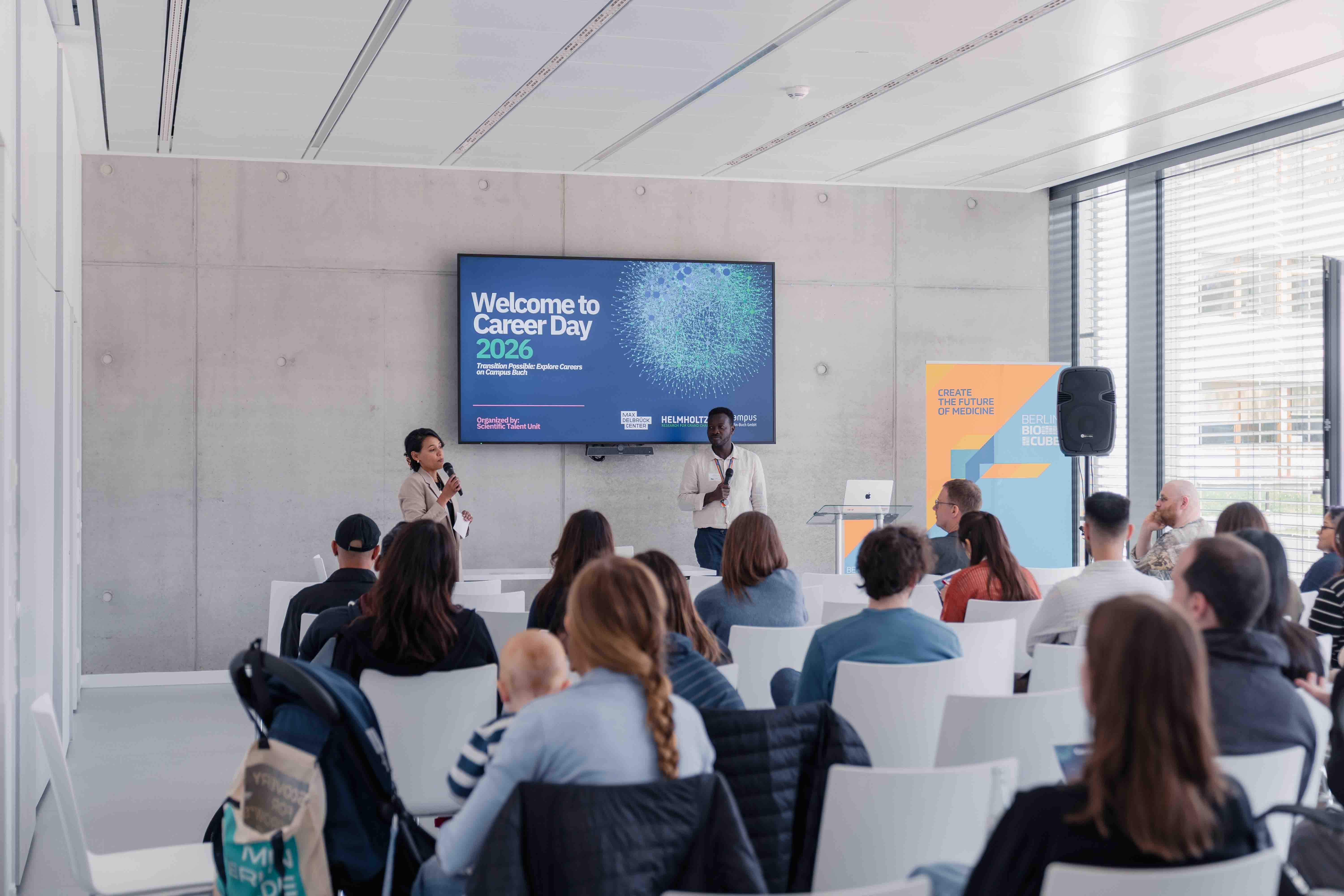
The Max Delbrück Center’s Career Day, held in cooperation with the operator of the Berlin-Buch Campus, offered young researchers insights into the working world of startups and biotech companies
April 16 was a packed day for participants in the Max Delbrück Center’s Career Day, titled “Transition Possible – Explore Careers Beyond R&D.” Numerous doctoral students and postdocs gained insights into potential careers outside the academic world.
The event was opened by biologist Lars Dittrich, who works as a science editor at MaiThink X. In the morning, there were virtual presentations across the Helmholtz Association on career paths outside of research. The speakers presented concrete entry-level and development opportunities and shared their experiences of transitioning from science to other professional fields.
In the afternoon, the Max Delbrück Center and Campus Berlin-Buch GmbH hosted an event at the BerlinBioCube startup center. Here, the Berlin BioScience Academy and the Innovation & Entrepreneurship Department of the Max Delbrück Center presented themselves. This was followed by lab tours at biotech companies and a workshop on the application process.
Pathways into the Biotech and Pharmaceutical Industries
How can you gain in-depth insights into the biotech and pharmaceutical industries without actually working in them? The Berlin BioScience Academy (BBA) offers exactly this opportunity. Its courses cover biotechnological and pharmacological processes, including Good Manufacturing Practice (GMP) and Good Clinical Practice (GCP). “Anyone considering a move into the industry or wanting to take the leap into a startup will gain an overview of the entire drug development process—from concept to market—in a very short time at the Biotech & Pharma Summer School,” said Dr. Uwe Lohmeier, who heads the BBA. Through its “Talk im Cube” event series, the BBA regularly brings together science and industry, offering panel discussions on topics such as financing strategies, female founders, CROs, and IP strategies in biotechnology. Here, too, participants have an easy opportunity to engage with biotech companies.
At the Max Delbrück Center, the Innovation & Entrepreneurship Department serves as a springboard for careers in spin-offs. Dr. Nevine Shalaby highlighted funding opportunities for future “sciencepreneurs” to develop innovative diagnostic or therapeutic approaches for practical application. The Innovation Office provides support through programs such as BOOST and PreGoBio to validate ideas and their basic feasibility, helps secure funding, offers mentoring, establishes contacts with industry and investors, and proactively supports business development.
How do biotech companies operate?
Over 50 biotech and medtech companies have set up their business in the BiotechPark Berlin-Buch, including numerous startups. Four of them opened their doors on Career Day to show participants how they work and what their mission is. T-knife, a spin-off of the Max Delbrück Center and Charité, introduced itself as a young biopharmaceutical company developing next-generation T-cell therapies to fight cancer.
CheckImmune, a spin-off of Charité, provided information about its work as an accredited specialized laboratory that supports the clinical development of new therapeutics through immunological studies, among other activities.
In the Biosynth laboratories, participants learned about the technologies used to develop and manufacture polymer-based excipients for drug delivery as well as bioconjugate drugs. Last but not least, the FyoniBio team presented its range of contract development and clinical laboratory services.
“There are surprisingly many different companies here,” said one participant, who could well imagine working at one of the biotech firms: “The labs in the BioCube are similar to those at research institutions, and the building feels very spacious, especially thanks to the large shared large common areas with a kitchen.” One of the participants noted with pleasant surprise how diverse the age range of employees in startups can be. Many found it fascinating to learn how the work culture in a startup functions and that the tasks there are different and more varied than in pure scientific research.
How successful is my application?
To wrap up the event, Career Day offered participants the chance to step into the shoes of a hiring manager during a group workshop. Anita Überheim, Head of Human Resources Europe at the global company Eckert & Ziegler SE, had the participants evaluate three anonymized CVs and cover letters and then explained which aspects matter in the selection process. She described how HR professionals proceed, how much time they have to review applications, which skills are important to mention, and what mistakes are common. Finally, the expert conducted a brief mock interview with one of the participants. During the joint evaluation with the audience, she explained how applicants should best communicate and respond. In addition to many helpful tips, a key insight for the young talents was this: It is not always necessary to meet 90 percent of the desired qualifications. What is far more important is that the person fits into the team and has the potential to continue developing.
The joint Career Day was very well received. “We are delighted to have had the opportunity to help organize this event. The Career Day offers concrete insights into the biotech industry and connects young talent with potential employers in the area, which is valuable for everyone involved,” says Dr. Ulrich Scheller, Managing Director of Campus Berlin-Buch GmbH.
Research, Patient care / 17.04.2026
A fundamentally new therapeutic approach to cystic fibrosis: Nanobody repairs cellular defect
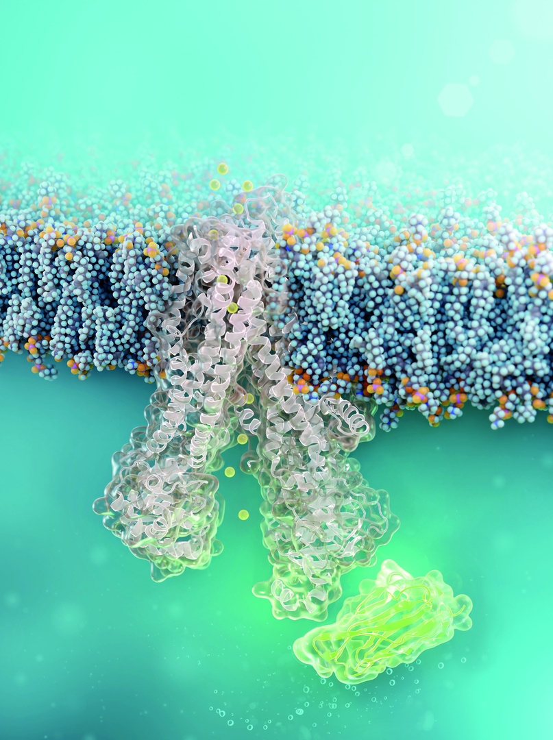
A tiny antibody component could fundamentally transform the treatment of cystic fibrosis: For the first time, researchers have succeeded in developing a so-called nanobody that penetrates directly into human cells and can repair the chloride channel most commonly affected in cystic fibrosis. The innovative therapeutic approach was developed in collaboration between teams from the Leibniz-Forschungsinstitut für Molekulare Pharmakologie (FMP) and Charité – Universitätsmedizin Berlin.The first authors of the study, which was recently published in the renowned journal Nature Chemical Biology, are Luise Franz (FMP) and Tihomir Rubil (Charité).
The clinical picture of cystic fibrosis—also known as CF—is caused by genetic defects in the so-called CFTR channel. This channel regulates water and salt transport in the lung mucosa and ensures the production of sufficiently fluid mucus.
In about 90 percent of cystic fibrosis patients, a mutation known as F580del is present in the CFTR channel, meaning that a single amino acid is missing at position 508 in its protein chain. This change causes CFTR to fold incorrectly and be broken down prematurely inside the cell, rather than functioning as a channel in the cell membrane of the airways. As a result, patients have thick mucus in their lungs, and pathogens can no longer be effectively cleared. The consequence is chronic infection and inflammation of the airways, leading to a progressive loss of lung function—in the worst-case scenario, this necessitates a lung transplant.
Professor Dr. Marcus Mall, Director of the Department of Pediatric Respiratory Medicine, Immunology and Critical Care Medicine at Charité, has, together with his team, made a significant contribution in recent years to noticeably improving the treatment of cystic fibrosis through therapy with three small-molecule drugs (CFTR modulators): With the help of the so-called triple therapy consisting of elexacaftor, tezacaftor, and ivacaftor (ETI), the function of the CFTR channel can be increased to about 50 percent of the normal level. However, chronic inflammation and infection of the lungs often persist, and there are also patients for whom this therapy is ineffective or whom cannot tolerate it.
An Antibody as a Repair Aid
There may be additional treatment options for this group in the future: The team led by chemist Professor Dr. Christian Hackenberger at the Leibniz-FMP has developed a new molecule in the lab that stabilizes the misfolded CFTR directly inside the cell. This is a nanobody—a tiny but stable antibody component that can bind precisely to defined surfaces of proteins. It is chemically modified with a “transport signal,” known as cell-penetrating peptides, which help it penetrate directly into the lung’s mucosal cells. There, the nanobody binds to the defective channel protein and helps it adopt the correct shape.
The researchers were able to demonstrate that the nanobody remained bound to the mutated CFTR channel in cells derived from cystic fibrosis patients for at least 24 hours. It did not damage the cells in the process. Functional studies also confirmed that the corrected channel once again transported chloride across the cell membrane.
Combination of triple therapy and nanobody
In combination with established ETI triple therapy, the nanobody demonstrates a pronounced synergistic effect in these cell cultures: While the ETI agents restored the function of the defective CFTR channel by about half on average, the channel activity could be increased to nearly 90 percent of normal levels through the additional administration of the nanobody.
The study thus demonstrates that exogenously administered cell-penetrating nanobodies can stabilize disease-relevant, misfolded proteins inside cells and restore their function. “In addition to the preclinical proof of concept for repairing the CFTR channel, this is the first example of a functional cell-permeable antibody: Until now, cell-permeable nanobodies have primarily been used to visualize intracellular target structures or for the targeted killing of cells,” says Prof. Dr. Christian Hackenberger.
“Since the nanobodies bind directly in the region of the F508del mutation, they enable even more targeted treatment of the maturation defect in CFTR channels,” says Prof. Dr. Marcus Mall. “This new mechanism of action allows CFTR function to be corrected significantly better in combination with existing CFTR modulators. Our results suggest that this new approach may even enable complete normalization of CFTR function. This would be another breakthrough for the treatment of cystic fibrosis.”
Thus, this work thus opens up new possibilities for further improving the treatment of cystic fibrosis—while also laying the groundwork for broader therapeutic applications.
Prospects beyond cystic fibrosis
However, key questions must still be resolved before the approach can be applied clinically to cystic fibrosis, such as developing a suitable formulation for inhalation and ensuring efficient penetration of the viscous CF mucus. Furthermore, it remains unclear how the nanobody acts within the body and how the immune system reacts to nanobody treatment. These challenges are currently being addressed within Collaborative Research Center 1449 “Dynamic Hydrogels at Biointerfaces,” within the framework of which the current results were also generated.
The approach of intracellular nanobody therapy could also be helpful beyond cystic fibrosis for other rare genetic diseases in which protein misfolding plays a role and for which there are currently few effective treatments.
About Cystic Fibrosis
Cystic fibrosis is one of the most common fatal hereditary diseases worldwide. As many as 8,000 children, teens, and adults are living with the disease in Germany today. An imbalance in salt and water levels in the body causes people with cystic fibrosis to produce thick, sticky secretions that harm organs such as the lungs and pancreas. This leads to progressive loss of lung function and shortness of breath, which still significantly lowers life expectancy despite advances in treatment. Some 150 to 200 children are born with this rare disease in Germany each year. A test for cystic fibrosis is part of routine screening for newborns.
Publication: Franz, L., Rubil, T., Balázs, A., Overtus, M., Kemnitz-Hassanin, K., Govaerts, C., Mall, M. A., & Hackenberger, C. P. R.. A cell-permeable nanobody to restore F508del cystic fibrosis transmembrane conductance regulator activity. Nature Chemical Biology 2026. doi: 10.1038/s41589-026-02199-w
Source: Joint Press Release from the Leibniz-Forschungsinstitut für Molekulare Pharmakologie and Charité – Universitätsmedizin Berlin
A fundamentally new therapeutic approach to cystic fibrosis: Nanobody repairs cellular defect
Innovation / 14.04.2026
Eckert & Ziegler: Metzler Initiates Coverage with a Buy Rating and a Price Target of € 21.00. Upside Potential: 41%
B. Metzler seel. Sohn & Co. AG (Bankhaus Metzler) has started research coverage of Eckert & Ziegler SE (ISIN DE0005659700), a leading supplier of isotope-based components for nuclear medicine and measurement technology, with a buy recommendation and a price target of € 21.00. This corresponds to an upside potential of 40.8% compared to the Xetra closing price of € 14.92 on April 13, 2026.
Bankhaus Metzler thus highlights the strong market position of Eckert & Ziegler SE, which is benefiting from rising demand for diagnostic and therapeutic radioisotopes in nuclear medicine.
About Eckert & Ziegler.
Eckert & Ziegler SE with more than 1,000 employees is a leading specialist for isotope-related components in nuclear medicine and radiation therapy. The company offers a broad range of services and products for the radiopharmaceutical industry, from early development work to contract manufacturing and distribution. Eckert & Ziegler shares (ISIN DE0005659700) are listed in the TecDAX index of Deutsche Börse.
Contributing to saving lives.
Quelle: https://www.ezag.com
Research, Patient care / 13.04.2026
First “protein map” of neurons that initiate pain
Helmholtz researchers have created the first detailed protein map of specific sensory neurons that trigger pain. Their study, published in “Nature Communications,” will help researchers better understand the molecular mechanisms of chronic inflammatory pain and identify new drug targets.
Joint press release by the Max Delbrück Center and the Helmholtz Centre for Infection Research
One in five people worldwide suffers from chronic inflammatory pain. Meanwhile, about two thirds of those affected find little relief from existing pain medications; new therapeutic approaches are urgently needed. “We first must understand precisely how sensory nerve cells trigger pain at the molecular level — in other words, which proteins are involved,” says Professor Gary Lewin, Group Leader of the Molecular Physiology of Somatosensory Perception lab at the Max Delbrück Center in Berlin.
To unravel these molecular processes, Lewin – who has been studying pain for four decades and recently discovered a previously unknown ion channel involved in pain perception – is working closely with systems biologist Dr. Fabian Coscia, Group Leader of the Spatial Proteomics lab at the same center. Coscia co-developed a method called Deep Visual Proteomics that makes it possible to determine the proteome — the complete set of proteins — of specific cells and to create maps detailing the spatial locations of individual proteins.
The researchers combined this technology with electrophysiological methods from Lewin’s group. This enabled them to first identify specific subtypes of pain neurons based on their function and then analyze their protein profiles. The result is a high-resolution molecular map of these nerve cells, which has been published in “Nature Communications.” The team also demonstrated how the technology can identify potential new drugs targets to treat chronic pain.
Dr. Sampurna Chakrabarti is the study’s first author and a former postdoctoral researcher in the Lewin lab who now heads the Pathways in Infection and Nociception group at the Helmholtz Centre for Infection Research in Braunschweig. Nociception refers to how our nerves respond to stimuli that trigger pain. Nerves in skin and other peripheral tissues – such as muscles and joints – that detect damaging stimuli are called nociceptors; they relay signals to the brain to initiate pain.
Undiscovered signaling pathways
All nociceptors are not alike. “Until now, only the transcriptome – that is, the RNA level information of the different subsets of nociceptors – was known,” says Chakrabarti. “However, the actual functional components of all cells are the proteins formed from these transcripts – and we have now examined them in greater detail for the first time in two subtypes of nociceptors.” Using an electrophysiological method known as the patch-clamp technique, the team first identified and characterized two nociceptor subtypes – peptidergic and non-peptidergic – in the spinal ganglia of mice. Each of these subtypes respond differently to similar stimuli and may initiate pain of different quality and duration.
The researchers used around 50 neurons of each subtype to generate a specific protein map for each of the two cell types. Deep Visual Proteomics combines mass spectrometry with microscopy, artificial intelligence and robotics. Coscia and his team have so far mainly used this methodology for proteome analyses of cancer cells. “We have now shown for the first time that it can also be applied to nerve cells,” he says.
The team measured more than 6,000 proteins in these 50 neurons. A comparison with existing RNA data revealed that the transcriptome and proteome of the cells differ significantly in some cases – an indication that key functional processes only become visible at the protein level. “We provide a unique molecular map of pain-initiating neurons,” says Coscia. “It enables the identification of signaling pathways in these cells that have so far remained hidden.”
In an additional step, Chakrabarti and her colleagues wanted to understand which proteins sensitize nerve cells, contributing to chronic pain. They isolated both types of nociceptors from mouse dorsal root ganglia and exposed them to a molecule called Nerve Growth Factor (NGF), which is known to trigger chronic pain both animals and humans, such as in arthritis. Using Deep Visual Proteomics, the researchers were able to precisely identify the proteins produced after the cells were exposed to NGF.
Reduced sensitivity to pain signals
Lewin and his team had already discovered that NGF plays an important role in chronic inflammatory pain more than 30 years ago. “In dogs and cats, pain can now be alleviated very effectively using antibodies that inhibit NGF,” says Lewin. “In humans, rare side effects have unfortunately prevented their use,” he adds. “But now we may have found an alternative approach: targeting a downstream protein responsible for NGF’s sensitizing effect.”
“We identified several proteins that were present in higher levels in a subset of nociceptors following treatment with NGF. The higher levels of these proteins could be linked to long term pain associated with inflammation,” says Chakrabarti. One of the proteins, an enzyme called B3GNT2, stood out in particular. “When we knocked out the corresponding gene in the cells, the inflammation-induced hyperactivity of nociceptors was reduced. Fewer cells responded to mechanical stimulus,” she says. In other words, the neurons had become less sensitive and would elicit much less pain.
In the future, the researchers plan to validate their findings in mice and humans. “More than 90 percent of all approved drugs now target proteins,” says Coscia. “This highlights how important it is to develop a better understanding of these molecules in order to identify new targets for more effective pain therapies and treatments for other neurological diseases.”
Text: Anke Brodmerkel
Further information
Molecular Physiology of Somatic Sensation
- Chakrabarti lab
- Profile of Gary Lewin
- Profile of Fabian Coscia
- Creating protein maps of tumors
- Understanding the roots of chronic pain
Literature
Sampurna Chakrabarti, Anuar Makhmut, Atena Mohammadi et al. (2026): “Deep visual proteomics uncovers nociceptor diversity and pain targets.” Nature Communications, DOI:10.1038/s41467-026 – 71418‑8
Innovation / 10.04.2026
Eckert & Ziegler Secures Patient Access to Critical Eye Cancer Treatment with MDR Certification for Ru-106 Eye Applicators
Eckert & Ziegler BEBIG GmbH, subsidiary of Eckert & Ziegler SE with focus on brachytherapy solutions for the treatment of eye tumors and prostate cancer, obtained the MDR certificate for its Ruthenium-106 (Ru-106) Eye Applicators from competent authorities. Eckert & Ziegler is the only global provider of these eye applicators. Therefore, this important milestone is a critical safeguard against treatment shortages.
The Medical Device Regulation (MDR) is a European Union directive (EU 2017/745) with the aim of improving the quality of medical devices and increasing patient safety. Obtaining this certification guarantees the long-term availability of Ru-106 Eye Applicators within the EU. These medical devices have been manufactured and internationally marketed by Eckert & Ziegler for more than 30 years. They are actively used in almost 50 countries and contribute several million euros in annual sales to the Eckert & Ziegler Group's earnings.
During an ophthalmic brachytherapy procedure, a small radioactive plaque containing Ru-106 is used to treat uveal melanoma in adults or retinoblastoma in children. The plaque is sutured to the wall of the eye, adjacent to the tumor, and left in place for several days until the required dose of radiation has been delivered. As an alternative to the removal of the affected eye, this treatment offers a chance for patients to conserve vision and quality of life.
"The dedication of our team to achieve MDR certification for a niche product like the Ru-106 Eye Applicators demonstrates our commitment to ensure the long-term availability of this vital form of therapy to treatment centers and patients," explained Katrin Antonenko, Managing Director of Eckert & Ziegler BEBIG GmbH. “The milestone furthermore marks the durable legacy of a product that started to pave the way for the sustainable and successful growth of the Eckert & Ziegler Group more than three decades ago. The experience gained during the successful approval process is expected to expedite additional projects.”
About Eckert & Ziegler
Eckert & Ziegler SE, with more than 1,000 employees, is a leading specialist in isotope-related components for nuclear medicine and radiation therapy. The company offers a broad range of services and products for the radiopharmaceutical industry, from early development work to contract manufacturing and distribution. Eckert & Ziegler shares (ISIN DE0005659700) are listed in the TecDAX index of Deutsche Börse.
Source: Press Release Eckert & Ziegler
Eckert & Ziegler Secures Patient Access to Critical Eye Cancer Treatment with MDR Certification for Ru-106 Eye Applicators
Research / 08.04.2026
Gilead acquires FMP and LMU Spin-off Tubulis and expands Oncology Pipeline with next-generation ADC
U.S. biopharmaceutical company Gilead has entered into a definitive agreement to acquire Tubulis GmbH. Tubulis was spun off in 2019 from the Leibniz Forschungsinstitut für Molekulare Pharmakologie (FMP) and LMU Munich and develops next-generation antibody-drug conjugates (ADCs) designed to deliver various active ingredients more selectively to tumors and maximize patient benefit. The transaction expands Gilead’s oncology pipeline with several innovative programs and platform technologies, some of which originated from academic research at the Leibniz Forschungsinstitut für Molekulare Pharmakologie (FMP) in Berlin.
The acquisition includes Tubulis’ lead product, TUB-040, a NaPi2b-targeted topoisomerase I inhibitor ADC currently in Phase 1b/2 clinical development for the treatment of platinum-resistant ovarian cancer and non-small cell lung cancer (NSCLC). Gilead will also acquire TUB-030, a 5T4-targeted ADC with promising early clinical data across various solid tumor types as well as Tubulis’ next-generation ADC platform and a promising early pipeline.
Dr. Dominik Schumacher, CEO and co-founder of Tubulis, also emphasizes the importance of combining scientific excellence with industrial development strength: „From the outset, we believed our conjugation technology platforms could have broad impact across the ADC field and the initial data from TUB-040 have reinforced that conviction,” said Dr. Dominik Schumacher, Chief Executive Officer and Co-founder of Tubulis. „Joining Gilead allows us to build on this foundation within an organization that brings deep scientific expertise, global development capabilities, and the scale needed to translate innovation into medicines for patients worldwide. Through our existing collaboration, Gilead has already seen the potential of our technologies and together, we are well positioned to accelerate the development of our ADC pipeline. I’m deeply grateful to the Tubulis team, our Board of Directors, investors, and partners for their commitment and helping make this milestone possible.”
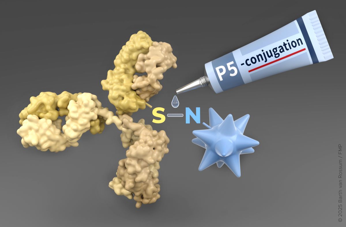
P5 Conjugation Technology from FMP
A key role in Tubulis’ product development is played by the innovative P5 conjugation technology, a linker chemistry that enables the precise coupling of antibodies with highly potent active ingredients. It was developed through basic research by Prof. Dr. Christian Hackenberger, one of Tubulis’ co-founders, and his team at the Leibniz Forschungsinstitut für Molekulare Pharmakologie (FMP). Tubulis has further developed P5 technology for clinical research and application in collaboration with the research group led by Prof. Dr. Heinrich Leonhardt (Ludwig Maximilian University of Munich), a co-founder of Tubulis. “This successful technological development demonstrates the potential of interdisciplinary collaboration across departmental and institutional boundaries”, says Prof. Dr. Heinrich Leonhardt. This technology forms a central platform for stable and controlled drug delivery in the company’s ADC programs. „This acquisition not only underscores the success of our conjugation technology and the performance of the Tubulis team in improving cancer therapy with new ADCs. It also demonstrates the importance and impact of basic research at universities and research institutes for translational innovation", says Prof. Dr. Christian Hackenberger.
Upon completion of the transaction, Tubulis will operate as an independent ADC research organization within Gilead, with the Munich site serving as a hub for ADC innovations. The company will build on existing integrated capabilities in research, production, and clinical development to advance next-generation ADCs.
Gilead will acquire all of the outstanding equity of Tubulis for 3.15 billion US Dollars in upfront cash consideration on a cash-free, debt-free basis, plus up to 1.85 billion US Dollars in contingent milestone payments. The acquisition is expected to close in the second quarter of 2026. Closing of the transaction is subject to expiration or termination of certain regulatory filings and other customary closing conditions.
About Tubulis
Tubulis develops tailored antibody-drug conjugates (ADCs) with improved biophysical properties. In preclinical models, the ADCs have already demonstrated targeted and sustained accumulation in the tumor as well as long-lasting anti-tumor effects. The two most advanced programs in the growing pipeline are TUB-040 (targeting NaPi2b) and TUB-030 (targeting 5T4). Both programs are currently being investigated in clinical trials for cancers with high unmet medical needs. For more information, visit: www.tubulis.com
Source: Leibniz-Forschungsinstitut für Molekulare Pharmakologie
Gilead acquires FMP and LMU Spin-off Tubulis and expands Oncology Pipeline with next-generation ADC
Research / 27.03.2026
Friedrich Stolz Award for the Tubulis Team
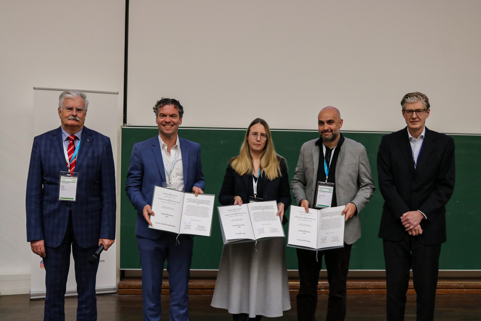
The Medicinal Chemistry Division of the Gesellschaft Deutscher Chemiker (GDCh) and the Pharmaceutical/Medicinal Chemistry Division of the Deutsche Pharmazeutische Gesellschaft (DPhG) have awarded the Friedrich Stolz Award 2026 to the team from Tubulis, a spin-off of the Leibniz-Forschungsinstitut für Molekulare Pharmakologie and LMU Munich. Honored are Prof. Dr. Christian Hackenberger from the Leibniz-FMP as co-founder and advisor and Dr. Jonas Helma-Smets, co-founder and CSO, Dr. Marc-André Kasper, VP Chemistry and Early Discovery and Dr. Annette Vogl, VP Biology and Translational Research, from Tubulis.
The Friedrich Stolz Award recognizes exceptional dedication, scientific discoveries, or innovative technologies with proven relevance that contribute to substantial therapeutic innovation or sustainable development and go beyond early research stages.
The Tubulis team was awarded for their P5 ethynylphosphonamidate conjugation chemistry, enabling a novel type of antibody-drug conjugates (ADCs).The conjugation chemistry, also termed P5-labeling, was developed at FMP in the research unit of Christian Hackenberger. This new ADC class is characterized by exceptional plasma stability, favorable pharmacokinetic properties, and pronounced anti-tumor efficacy. Building on this, NaPi2b-targeting exatecan ADCs with particularly stable linkers, reduced off-target toxicity, and optimized molecular architecture were generated. The culmination of this work is the development candidate TUB-040, which in a clinical Phase 1 study in patients with platinum-resistant ovarian cancer demonstrates promising tumor remissions, a wide therapeutic window, and good tolerability. This work shows how innovative chemistry can be directly translated into novel, potentially more effective treatment options for hard-to-treat cancers.
The prize is awarded in memory of industrial pharmacist and drug researcher Friedrich Stolz, whose visionary innovative power serves as a model for application-oriented, therapeutically relevant research.
Photo: Awarded the Friedrich Stolz Prize: Prof. Dr. Stefan Laufer (GDCh), Prof. Dr. Christian Hackenberger, Dr. Annette Vogl, Dr. Jonas Helma-Smets and Dr. Franz von Nussbaum (from left to right). © GDCh
Source: FMP
Friedrich Stolz Award for the Tubulis Team
Innovation / 26.03.2026
Eckert & Ziegler: Strong FY 2025 with Positive Outlook
Eckert & Ziegler SE (ISIN DE0005659700, TecDAX) set a new record in fiscal year 2025 with sales of €312.0 million. Compared to the previous year, sales rose by approximately €16 million (+5%). EBIT before special items from continuing operations (adjusted EBIT) increased by just under €12 million year-over-year to €77.7 million (+18%). Net income rose by approximately €15 million (+46%) to €48.8 million; this corresponds to earnings per share of €0.78 (previous year: €0.53, adjusted for stock split).
In the Medical segment, sales increased by €22.6 million, or 15%, to €171.3 million. The business with pharmaceutical radioisotopes remains the most important source of revenue.
The Isotope Products segment generated sales of €150.1 million, a decrease of €7.9 million (-5%) compared with the previous year. This was mainly due to a temporary shift toward lower-margin products compared with the same period last year. In addition, the cyberattack in 2025 led to project-related delays.
For the 2026 fiscal year, the Executive Board expects an adjusted EBIT of approximately €80 million. The corresponding sales forecast amounts to approximately €320 million. This forecast is based on a weighted average exchange rate of $1.20 per euro. Adjusted for currency effects and the licensing business, this corresponds to a growth of 9% in sales and 21% in earnings (adjusted EBIT).
The Executive Board and Supervisory Board will propose to the Annual General Meeting a dividend of €0.22 (previous year: €0.17, adjusted for stock split) per share entitled to dividends.
The 2025 financial statements can be found here: https://www.ezag.com/fy2025en/
Overview
FY 2025:
- Sales: €312.0 million (previous year: €295.8 million)
- adjusted EBIT: €77.7 million (previous year: €65.9 million)
- Net income: €48.8 million (previous year: €33.3 million)
Forecast 2026:
- Sales of approx. €320 million
- adjusted EBIT of approx. €80 million
About Eckert & Ziegler.
Eckert & Ziegler SE with more than 1.000 employees is a leading specialist for isotope-related components in nuclear medicine and radiation therapy. The company offers a broad range of services and products for the radiopharmaceutical industry, from early development work to contract manufacturing and distribution. Eckert & Ziegler shares (ISIN DE0005659700) are listed in the TecDAX index of Deutsche Börse.
Contributing to saving lives.
Research, Innovation, Patient care / 23.03.2026
Launch of the Einstein Center for Early Disease Interception
At the Einstein Center for Early Disease Interception, researchers from twelve Berlin institutions will be pooling their expertise to explore new approaches to prevention, long before symptoms appear, and bring them quickly into practice. The center opens today with a ceremony
Joint press release by the Max Delbrück Center, Charité – Universitätsmedizin Berlin, Berlin Institute of Health at Charité, and Technische Universität Berlin
Serious diseases often develop unnoticed over many years. By the time symptoms appear, organ damage is often too extensive to be reversed completely. In many cases, there are no effective treatments to stop diseases from progressing.
Researchers from twelve leading Berlin institutions are heading the charge to change the way we think about prevention. Using the latest technologies – including advances made by researchers from Berlin – they aim to unravel what happens inside cells at the very earliest stages of disease. Such knowledge will enable them to develop early interventions, when only individual cells are affected and diseases are still controllable.
The Einstein Center for Early Disease Interception (EC-EDI) is bringing together the necessary expertise to study such early disease mechanisms. The center is being officially inaugurated today at the Max Delbrück Center’s Berlin Institute for Medical Systems Biology in Mitte after a two-year preparatory phase. Invited guests include politicians, scientists and the public. The Einstein Foundation Berlin is funding the center with €6 million and is supporting the recruitment of young international talent with additional funds from the state of Berlin.
A dynamic network for key technologies
Over the next six years, researchers will further develop, integrate, and apply key technologies to the effort. These include single-cell multiomic and spatial biology technologies, patient-specific organoids, 3D bioprinting, and AI-based modeling of mechanisms and disease trajectories. A platform for cross-institutional collaboration will also be established to help translate research findings into clinical practice more quickly in partnership with industry and investors – while generating societal and economic value for the state of Berlin. Initial focus areas include respiratory and neurological diseases, such as inflammatory lung diseases, tuberculosis, Alzheimer’s disease, and multiple sclerosis.
Participants in EC-EDI include Charité – Universitätsmedizin Berlin, the Berlin Institute of Health at Charité, the Max Delbrück Center, Technische Universität Berlin, Freie Universität Berlin, Humboldt-Universität zu Berlin, and additional non-university institutions such as the Museum für Naturkunde and several Max Planck Institutes.
Statements from Einstein Center spokespersons
Professor Nikolaus Rajewsky (Head Spokesperson; Director of the Berlin Institute for Medical Systems Biology at the Max Delbrück Center and Professor at Charité):
“We are advancing molecular prevention and intervention – and aim to bring our research to patients as quickly as possible. Major breakthroughs do not arise solely in the clinic, in labs, or on a computer. The Einstein Center creates a shared, open platform with clear rules and short pathways. Clinicians, basic researchers, and data scientists will collaborate across institutions, as well as with companies and investors. This will enable us to work faster and will hopefully help further establish Berlin as a leading international hub in this field.”
Professor Leif Erik Sander (Project Head; Director of the Department of Infectious Diseases and Critical Care Medicine at Charité and Research Group Leader at BIH):
“Conventional methods often fail to detect the early signs of emerging diseases. Moreover, we don’t always understand what is actually going wrong at the molecular level or how we could intervene to stop these diseases. This is where early disease interception comes in: Using high-resolution technologies, we aim to understand what sets things on the wrong track and create opportunities to correct that course very early on. The train, so to speak, needs to be put back on the track to health. We want to use our findings to develop innovative solutions, such as new diagnostic tests and medications. This is how research leads to medical progress and value creation – and that strengthens Berlin as a hub for innovation and business.”
Dr. Janine Altmüller (Head of the Core Unit Genomics at the Berlin Institute of Health at Charité, BIH):
“New methods are key to the Einstein Center’s vision: Never before has it been possible to examine characteristic features such as nucleic acids or proteins of individual cells in their natural tissue context with such precision and high resolution. Even the smallest changes that mark the onset of a disease can now be digitalized and, with the help of AI for example, better deciphered and understood. This knowledge is crucial for developing targeted interventions. At the Einstein Center, experts from various disciplines will be working together toward this goal, thereby accelerating the translation of scientific findings into clinical applications.”
Professor Jens Kurreck (Executive Director of the Institute of Biotechnology, Technische Universität Berlin):
“The Einstein Center for Early Disease Interception provides a unique platform for close collaboration among leading Berlin-based research institutions and enables a better understanding of the early mechanisms of disease development using various human models. It is particularly important to offer young scientists access to state-of-the-art technologies within this consortium and to create optimal conditions for them to build their scientific careers.”
Further information
Research, Innovation, Patient care / 19.03.2026
Strengthen international cooperation in clinical trials
As part of the Australian Clinical Trials Initiative, ACTI, Australian companies and representatives of the Australien Government’s Trade and Invest Commission, Austrade, visited the Berlin-Buch Science and Biotech Campus today. The initiative promotes Australia as an attractive location for clinical trials.
Dr. Christina Quensel, CEO der Campus Berlin-Buch GmbH, welcomed the guests and, during a tour, provided information about the campus’s ecosystem of basic biomedical and clinical research, life science startups, and biotechnology companies. Biotech and medtech companies operate here in one of Europe's leading biotech parks.
The delegation gained insight into two scientific core facilities the Proteomic Lab of the Berlin Institute of Health in the Charité und the Cryo Electron Microscopy, a collaborative scientific infrastructure involving Charité – Universitätsmedizin Berlin, the Max Delbrück Center, and the Leibniz Research Institute for Molecular Pharmacology.
At the BerlinBioCube startup center, the guests visited the startups CheckImmune GmbH, a CRO, and T-knife GmbH, a pharmaceutical company that develops highly innovative cancer therapies.
During a networking lunch, the delegation engaged in intensive discussions with executives and stakeholders of the BiotechPark Berlin-Buch.
Many thanks to our guests for the exciting exchange!
Minesh Lalla, Senior Business Development Manager at Peter MacCallum Cancer Centre (CTS)
Jason Erasmus, Business Development Manager at Beyond Drug Development (CRO)
Tim Vale, Business Development Manager at Resolian Bioanalytics (Lab)
Karl Boehle, Senior Business Development Manager, Austrade
Pallavi Mishra, South Australia Trade & Investment Manager, Austrade
George Zafiris MBA, Prime Financial Group (R&D Tax Advisors)
Bellberry (Ethics)
The delegation then proceeded to Berlin Partner for Technology and Business, which, in cooperation with Austrade, had hosted an exclusive seminar on opportunities for German-Australian collaboration in clinical trials. The event highlighted how the Australian clinical trial ecosystem is structured and the key strengths and efficiency advantages it offers. Executives were invited The event was aimed at pharmaceutical and biotech R&D leaders, clinical operations and trial strategy decision-makers, CROs and clinical service providers, as well as leaders from academic, hospital, and research institutions, and stakeholders in policy, innovation, and the ecosystem.
Research / 17.03.2026
Fat tissue fuels gut inflammation
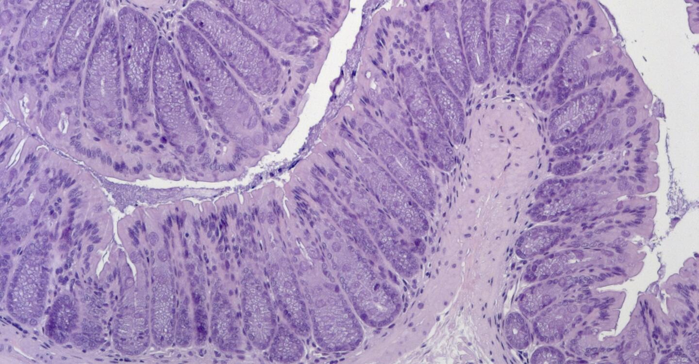
Researchers at the Max Delbrück Center and Charité – Universitätsmedizin Berlin show that adipose tissue amplifies gut inflammation in Crohn’s disease. The study, published in the “Journal of Clinical Investigation,” also reveals how genetic changes in immune cells may contribute to the disease.
Crohn’s disease is a chronic inflammatory disorder of the intestinal tract affecting millions of people worldwide. One prominent feature of the disease is “creeping fat” — adipose tissue that wraps around inflamed sections of the intestine. Scientists have long suspected that this fat tissue may influence disease progression, but the underlying mechanisms have remained unclear
Now, researchers from Charité – Universitätsmedizin Berlin, the Berlin Institute for Medical Systems Biology at the Max Delbrück Center (MDC-BIMSB) and their collaborators show that fat tissue releases signals that ultimately damage the intestinal barrier in Crohn’s disease. They also show that acquired genetic mutations in immune cells may fuel the disease.
“There is a longstanding link between obesity and Crohn’s disease,” says Dr. Ashley Sanders, Group Leader of the Genome Instability and Somatic Mosaicism Lab at the Max Delbrück Center and a senior author of the paper. “While it’s fairly well established that adipose tissue secretes molecular signals that promote inflammation, here we detail the link between fat-derived signals and intestinal inflammation.”
Mice with no fat tissue
The researchers first conducted experiments in a mouse model of generalized lipodystrophy – an extremely rare disease in which affected people lack fat tissue among other symptoms.
When they exposed the mice to a chemical substance known to cause intestinal inflammation, they found the lipodystrophy mice were more resistant than normal mice. Their intestinal barrier remained stable and they had low levels of pro-inflammatory immune cells, particularly Th1 and Th17 T cells – specialized subsets ofimmune cells that coordinate immune responses.
When the team transplanted adipose tissue back into the lipodystrophy mice, they became more susceptible to the chemically induced gut inflammation. Remarkably, they observed this effect only when the transplanted fat was capable of producing leptin, suggesting that leptin plays a central role in regulating the immune system in mice.
“These findings were expected because previous research had already established that fat tissue is an active immune organ,” says Dr. Britta Siegmund, a senior author of the paper at Charité — Universitätsmedizin Berlin. “But they nevertheless highlight that signals derived from adipose tissue can substantially amplify intestinal inflammatory responses.”
Puzzling clinical findings
However, the results appeared to conflict with findings from a clinical case at Charité: A patient with acquired generalized lipodystrophy who had developed Crohn’s disease, despite having no fat tissue at all. “This was curious and we wanted to understand why this patient had developed the disease,” Siegmund adds.
Using single-cell sequencing, the Sanders lab examined immune cells extracted from the patient’s blood and intestinal tissue. The analyses showed an unusually large group of cells that carried a mutation in a gene called NRAS, which helps regulate cell growth and survival. This suggested that these cells had multiplied from a single original cell in a process called clonal expansion – such mutated cells arise spontaneously and are not inherited. Cells with mutated NRAS survive longer and divide more than they should, potentially driving chronic inflammation.
“This mutation likely helped the inflammatory T cells survive and expand over time,” explains Sanders. “That persistent expansion may have fueled the systemic and intestinal inflammation that was observed. What’s more, our results show how powerful single-cell genomic technologies can be for uncovering hidden genetic changes in individual immune cells,” she adds.
Toward more personalized treatment
Among other pro-inflammatory signals, adipose tissue also secretes mediators called adipokines — including leptin, which is best known for regulating appetite and metabolism. In the study, the lipodystrophy patient was given a synthetic form of leptin to treat type-2 diabetes. While the hormone improved the patient’s metabolic health, it also increased pro-inflammatory signaling and raised levels of Th17 immune cells.
“The finding highlights how adipokines such as leptin and genetic changes in immune cells interact to influence chronic inflammatory diseases,” says Dr. Carl Weidinger, another senior author of the study at Charité. This dual role of metabolic and genetic factors could help explain why Crohn’s disease behaves differently in different people, he adds. “Understanding these interactions may help us develop more precise, personalized approaches to treatment.”
Text: Gunjan Sinha
Further information
Genome instability and somatic mosaicism
Literature
Marilena Letizia, Toka Omar, Patrick Weidner et al. (2026): “Characterization of intestinal immune responses in generalized human and murine lipodystrophy.” Journal of Clinical Investigation, DOI: 10.1172/JCI192322.
Image
Colon tissue from a mouse model of acquired generalized lipodystrophy. Colon tissue remains intact after exposure to a chemical that causes gut inflammation. © Anja Kühl, Charité
Research, Patient care / 13.03.2026
How cancer immunotherapies affect the heart
Checkpoint inhibitors help fight cancer but increase the risk of heart disease. An interdisciplinary team, including researchers from the Max Delbrück Center, is investigating how the therapy can be made safer. The Hector Foundation is supporting the project with €1.3 million.
Checkpoint inhibitors have become standard cancer therapy in recent years. These antibody drugs stimulate the immune system to attack malignant cells more aggressively. They were first widely used to treat patients with melanoma, but are now also indicated for patients with lung, kidney, or bladder cancer because the immunotherapy treatment is more effective than other options.
However, the drugs are not good for the heart. “We estimate that cancer patients receiving immunotherapy with checkpoint inhibitors have about a threefold increased risk of heart disease such as coronary artery disease,” says cardio-oncologist Dr. Lorenz Lehmann from Heidelberg University Hospital. Together with an interdisciplinary team of researchers, Lehmann is studying the biological mechanisms that cause these side effects in an effort to reduce the therapy’s impact on the heart.
The consortium, called ATHERIC-CARE (Atherosclerosis Risk in Cancer Patients after Immune Checkpoint-Inhibitor Therapy – Assessment and Exploration), includes Dr. Isabel Poschke from the German Cancer Research Center in Heidelberg, Dr. Henrike Maatz from the Max Delbrück Center in Berlin-Buch, Professor Sylvia Saalfeld from Kiel University, and Professor Oliver Müller from the University Hospital Schleswig-Holstein in Kiel. Beginning February 1, 2026, the Hector Foundation will fund the multicenter team’s research with €1.3 million over three years.
Inflamed coronary arteries
Coronary artery disease is a form of atherosclerosis affecting the coronary arteries. Lipids and other substances from the bloodstream accumulate on the inner walls of the vessels and trigger chronic inflammation. Over time, the blood vessels become increasingly narrowed, which can lead to heart failure or a heart attack. ATHERIC-CARE is primarily investigating how checkpoint inhibitors activate or accelerate this inflammatory mechanism.
The scientists are using DNA analyses to investigate which immune cells drive atherosclerosis, for example. In animals, they are studying early inflammatory mechanisms and analyzing computed tomography images of affected individuals with assistance from machine learning tools. Such analyses can not only detect metastases, but also reveal changes in the coronary arteries.
“Our task will be to determine which genes become active in the cells of the coronary arteries and in the surrounding tissue – in other words, what exactly is happening at the molecular level in the different cell types,” says Dr. Henrike Maatz, a scientist in the Genetics and Genomics of Cardiovascular Diseases lab headed by Professor Norbert Hübner at the Max Delbrück Center. To find out, she will primarily be using two methods: single-nucleus RNA sequencing (snRNA-seq) and spatial transcriptomics.
Research for better quality of life
“For example, we want to characterize more precisely the interaction between immune cells and endothelial cells, which line the inside of blood vessels,” explains Maatz. “Our goal is to understand how immunotherapy promotes T cell-mediated inflammation in blood vessels, which can lead to unstable vascular deposits and atherosclerosis.” Using spatial transcriptomics, she also aims to determine how immune cells are distributed within plaques. The Hector Foundation is supporting the research with about €286,000.
“By bringing together the results generated in the consortium’s different research areas, we aim to identify biomarkers and strategies that make immunotherapy safer – and thereby improve patients’ quality of life,” says project leader Lehmann.
Maatz is looking forward to the upcoming collaboration: “Together, we will of course achieve much more than a single research group could,” she says. “And we have already worked very successfully with Lorenz Lehmann on other projects.”
Further information
Research / 07.03.2026
Equality in research drives innovation
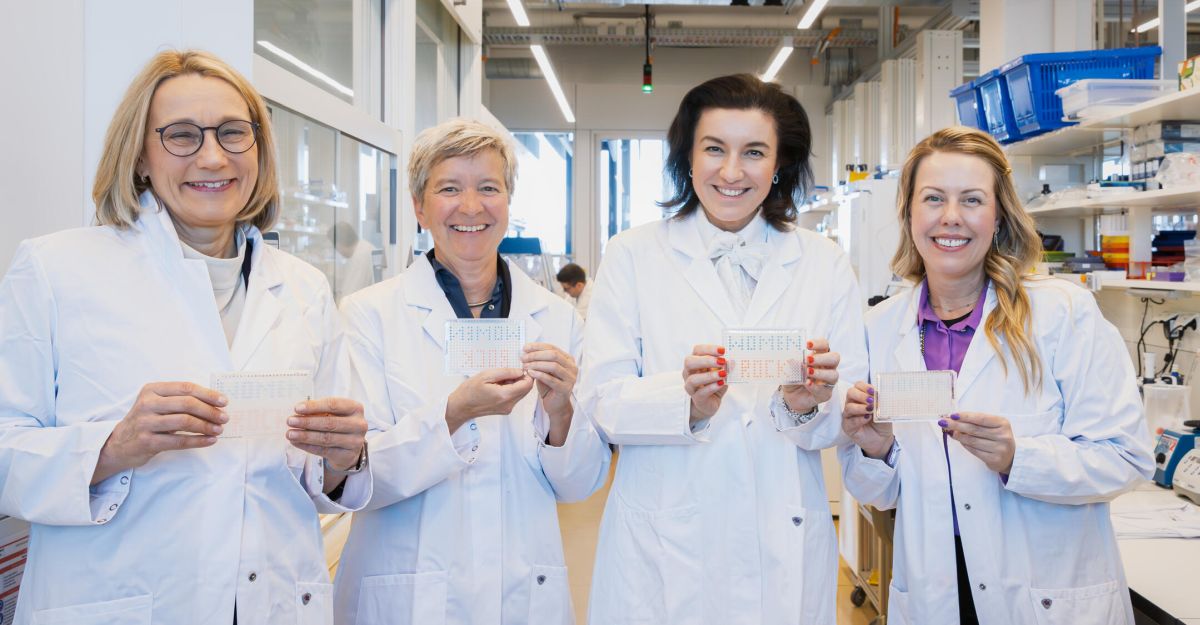
Marking International Women's Day, Federal Research Minister Dorothee Bär met with female researchers at the Max Delbrück Center
Statement from Federal Research Minister Bär:
“Equality is essential for Germany's capacity for innovation and long-term competitiveness. It is a decisive driver of progress. If we want to address the major challenges and megatrends of our time, be it digitalization, space research, medicine of the future, or key technologies, we cannot afford to leave half of the brightest minds out of driving Germany’s future forward. This is why the Federal Ministry of Education and Research, together with the states, is supporting equality at universities with a 2030 female professors program and the visibility of female researchers with the “Focus on Innovative Women” funding guidelines. In order to further strengthen social awareness of gender issues in research, we are currently designing a federal scientific award for gender research, which is to be announced later this year.”
Statement from Professor Maike Sander, Scientific Director and Chair of the Executive Board of the Max Delbrück Center:
“The discoveries made at the Max Delbrück Center lay the foundation for the medicine of the future. To get there, we need talented individuals who are enthusiastic about innovation and eager to contribute their ideas – regardless of gender or background. A total of 60 percent of our researchers are women. We have also been successful in recruiting early-career female group leaders. However, we still have some catching up to do when it comes to permanent professorships. This is why we specifically promote female scientists, for example through mentoring programs. We also approach exceptional candidates worldwide. More than half of our group leaders have been recruited internationally. We want to create an inspiring environment for all of them. We need a scientific culture in which careers are defined not by traditional role expectations, but by curiosity, talent, and courage. There is still much to be done.”
Photo: From left to right: Maike Sander, Katja Simon, Dorothee Bär and Ashley Sanders in a lab.
Maike Sander is Scientific Director of the Max Delbrück Center. Katja Simon is Head of Scientific Talent Management, Dorothee Bär is Federal Minister of Research, Technology and Space, Ashley Sanders is scientific group leader.
© Felix Petermann, Max Delbrück Center
Source: Max Delbrück Center
Equality in research drives innovation
economic development, Innovation / 03.03.2026
T-knife Therapeutics Announces Authorization of Clinical Trial Application for TK-6302, A Multi-Armored, CRISPR based T cell Therapy for Solid Tumors
Phase 1 ATLAS trial to evaluate TK-6302 in patients with advanced PRAME-positive solid tumors authorized for initiation
SAN FRANCISCO and BERLIN, March 03, 2026 - T-knife Therapeutics, Inc., a biopharmaceutical company developing next-generation T cell therapies to fight cancer, today announced the authorization of its Clinical Trial Application (CTA) to begin the Phase 1 ATLAS trial of TK-6302 in Europe. TK-6302 is a multi-armored PRAME-targeted T cell therapy specifically engineered to overcome the challenges associated with treating solid tumor cancers. The ATLAS trial, which is poised to begin this year, is an adaptive, first-in-human, open-label, Phase 1 trial of TK-6302 in patients with advanced PRAME-positive solid tumors.
“We are proud to achieve this important milestone, which enables the initiation of the Phase 1 ATLAS clinical trial and our transition to becoming a clinical-stage company,” stated Thomas M. Soloway, President and Chief Executive Officer of T-knife. “TK-6302 is a highly differentiated therapy, engineered for greater potency against PRAME, a clinically validated target with attractive commercial potential. PRAME is highly prevalent across multiple high unmet-need solid tumor indications, including squamous non-small cell lung, ovarian, endometrial, skin, and triple-negative breast cancers. Today’s announcement reflects the dedication, expertise, and urgency our team brings to transforming innovative science into life-changing therapies.”
Peggy Sotiropoulou, Ph.D., Chief Scientific Officer of T-knife, added, “The CTA was supported by a comprehensive and compelling preclinical data package demonstrating TK-6302’s best-in-class anti-tumor efficacy. By bolstering T-cell fitness and persistence while overcoming challenging tumor mediated immune barriers, TK-6302 has the potential to deliver meaningful clinical benefit, including deep, durable responses across a range of solid tumor cancers. Bringing a first-of-its-kind multi-armored, CRISPR gene-edited T cell therapy into the clinic underscores our commitment to pushing scientific boundaries in the service of delivering transformative therapies to patients.”
About TK-6302
TK-6302 is a PRAME-targeted T cell therapy that has been “supercharged” by the inclusion of multiple armoring innovations: a high affinity PRAME targeting receptor to enhance cytotoxicity; a costimulatory CD8 coreceptor to engage CD4 T cells and enhance T cell fitness and persistence; and a FAS-based checkpoint converter designed to boost engraftment and promote T cell survival in the hostile tumor micro-environment. Preclinical data with TK-6302 demonstrated sustained serial killing and cytokine secretion in a model mirroring the inhibitory ligand expression in PRAME-expressing tumors. In a complex 3-dimensional (3D) spheroid tumor model, TK-6302 eliminated multiple rounds of tumors and demonstrated superior anti-tumor activity compared to controls. TK-6302 is manufactured with a non-viral gene editing process for improved T cell receptor expression, and it has been successfully manufactured at-scale using the clinical process.
About T-knife Therapeutics
T-knife is a biopharmaceutical company dedicated to developing T cell therapies to deliver broad, deep and durable responses to solid tumor cancer patients. The company’s unique approach leverages its proprietary platforms and synthetic biology capabilities to design the next-generation of supercharged T cells with best-in-class potential.
T-knife was founded by leading T cell and immunology experts utilizing technology developed at the Max Delbrück Center for Molecular Medicine together with Charité – Universitätsmedizin Berlin, is led by an experienced management team, and is supported by a leading group of international investors, including Andera Partners, EQT Life Sciences, RA Capital Management and Versant Ventures. For additional information, please visit the company’s website at www.t-knife.com.
www.t-knife.comResearch / 25.02.2026
Gut microbes affect the heart via the brain
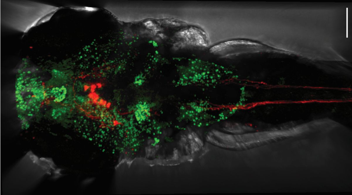
Researchers at the Max Delbrück Center have uncovered how a bacterial metabolite produced in the gut regulates heart function through specific brain cells. The findings, published in “Circulation Research,” reveal a new gut-brain-heart axis and point to potential therapies for hypertension and heart failure.
Hypertension and heart failure affect millions worldwide. Yet in many patients, doctors cannot fully explain why the heart becomes stiff and struggles to relax – a condition known as diastolic dysfunction.
Researchers in the lab of Dr. Suphansa Sawamiphak, Group Leader of Cardiovascular-Hematopoietic Interaction at the Max Delbrück Center, have identified a direct communication line between gut bacteria, the brain, and the heart. Using zebrafish as a model, the team discovered that certain gut microbes produce a small molecule called indole-3 acetic acid (IAA) from the dietary amino acid tryptophan. IAA acts on neurons in the brain, which in turn, control the heart. The study was published in “Circulation Research.”
“We were surprised that a single bacterial metabolite could influence the central nervous system, the heart, and major hormonal systems at the same time,” says Bhakti Zakarauskas-Seth, lead author of the paper. “It shows that the brain can act as a central hub in gut-heart communication.”
Tracking a signal from gut to brain
To understand how gut bacteria might influence the heart, the researchers focused on a distinct group of neurons in the hypothalamus known as hypocretin (Hcrt) in zebrafish larvae. These cells produce Hcrt neuropeptides, also known as orexins, which regulate many involuntary functions in the body – such as sleep and hunger – and also heart activity. When IAA levels dropped, Hcrt neurons became overactive. This increased sympathetic nerve signals to the heart, causing the heart muscle to stiffen, impairing its ability to relax properly.
When the researchers supplemented the larvae with IAA, neuronal activity normalized, heart function and blood pressure improved, and even related hormones such as renin and angiotensinogen returned to healthier levels.
They then examined data from a cohort of patients – humans also have Hcrt neurons – and found that IAA levels were reduced in patients with hypertension. Notably, they observed a sex-specific effect, with hypertensive women showing significantly lower levels of IAA in their serum samples than men.
Implications for patients and prevention
Diastolic dysfunction very common – up to half of all people over age 70 have some level of impairment. It is also is the underlying functional mechanism of heart failure with preserved ejection fraction (HFpEF), which accounts for over 50% of all heart failure cases.
For these patients, the findings open several potential avenues for better care, says Zakarauskas-Seth. “IAA levels could serve as a biomarker to identify patients at high risk of hypertension or heart failure. Therapeutically, boosting IAA production – for example through diet, probiotics, or supplementation – could become a new strategy to prevent or treat cardiovascular disease.”
That a single bacterial metabolite can influence the central nervous system, the heart, and major hormonal system also underscores a broader message, she adds. “The body does not operate in isolated compartments. Gut health, microbial balance, and diet directly shape how well the heart functions.” The researchers will need to validate their findings in other animal models and clinical studies will be needed to determine whether restoring IAA can benefit patients.
Text: Gunjan Sinha
Photo: Rückenansicht einer Zebrafischlarve: Aktive Nervenzellen sind grün, Hypocretine herstellende Neurone und ihre Ausläufer rot dargestellt. © Bhakti Zakarauskas-Seth, AG Sawamiphak, Max Delbrück Center
Source: Press Release Max Delbrück Center
Gut microbes affect the heart via the brain
Research / 24.02.2026
Similar kinases play distinct roles in the brain
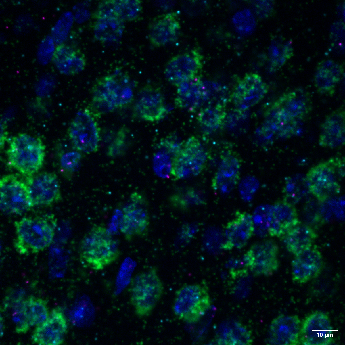
Researchers at the Max Delbrück Center show that two closely related enzymes, MNK1 and MNK2, act on the brain to regulate different behaviors. Their findings, published in “Molecular Psychiatry,” could help guide development of more precise therapies for neurological disorders.
Structurally, they look similar: MNK1 and MNK2 belong to the same enzyme family and are best known for regulating how cells make proteins. Their starring role in such a crucial cellular function has cast them into the spotlight as potential drug targets to treat nervous system disorders and chronic pain. But would it matter whether a drug targets only one of them?
In a study published in “Molecular Psychiatry,” researchers led by Rosalba Olga Proce, a doctoral student in the Molecular and Cellular Basis of Behavior lab led by Dr. Hanna Hörnberg at the Max Delbrück Center, set out to determine whether the two enzymes – also called kinases – perform distinct functions in the brain. The team found clear differences. Mice lacking MNK1 showed less interest in newly introduced objects than controls and impaired memory of objects. By contrast, mice without MNK2 appeared normal in object recognition tests but showed enhanced interest in social contacts.
“The behavioral differences we observed suggest that each kinase has a specialized function,” says Proce. “It might be preferable to target each kinase individually when designing drugs.”
Mouse behavior and molecular data
To uncover how these behavioral changes arose, the researchers used genetically engineered mice in which either the MNK1 or MNK2 gene was knocked out. They then combined behavioral testing with large-scale molecular analyses of brain tissue.
The team focused in particular on the cerebral cortex and on synapses – contact points where neurons communicate. Using proteomics, transcriptomics, and phosphoproteomics, they compared protein levels, gene expression, and protein phosphorylation patterns between the two types of mice.
Mice lacking MNK1 had an abundance of ribosomal proteins, which play a central role in transcribing messenger RNA into proteins. By contrast, in mice lacking MNK2, the expression and phosphorylation of proteins responsible for transmitting messages in synapses was reduced.
Notably, the researchers found that the molecular differences between the two kinases were far more pronounced in synapses than in whole-brain samples.
“We were surprised to see these differences,” says Hörnberg. “This suggest that the kinases may have different functions in the cell body compared to synapses.”
Toward more selective therapies
Instead of broadly inhibiting both kinases, future therapies might selectively target one kinase or the other to achieve more specific effects, Hörnberg adds.
She and her colleagues now aim to identify other molecules with which MNK1 and MNK2 interact. They also plan to study in greater detail, how these kinases regulate translation of messenger RNA into proteins during synaptic plasticity – the strengthening or weakening of connections between neurons over time. Synaptic plasticity is the fundamental mechanism underlying learning and memory – enabling the brain to rewire itself based on experience.
By clarifying how protein synthesis is fine-tuned at synapses, the team hopes to lay the groundwork for more precise interventions in neurological and psychiatric disorders.
Text: Gunjan Sinha
Photo: Section of mouse brain tissue showing that messenger RNA for MNK1 (magenta) and MNK2 kinase (turquoise) – important enzymes that regulate the production of proteins – are both expressed in excitatory neurons (green) in the cortex.
© Erika Uddström, Hörnberg Lab, Max Delbrück Center
Source: Press Release Max Delbrück Center
Similar kinases play distinct roles in the brain
Research / 18.02.2026
New material boosts MRI image quality
MRI scans often struggle to capture clear images of deep or delicate tissues. Researchers at the Max Delbrück Center now report in “Advanced Materials” a new lightweight antenna that boosts image quality and may shorten scan times ¬– without changing existing MRI machines.
Magnetic resonance imaging (MRI) is one of medicine’s most powerful diagnostic tools. But certain tissues deep inside the body – including brain regions and delicate structures of the eye and orbit that are of particular relevance for ophthalmology – are difficult to image clearly. The problem is not the scanner itself, but the hardware that sends and receives radio signals.
Now, researchers led by Nandita Saha, a doctoral student in the Experimental Ultrahigh Field Magnetic Resonance lab of Professor Thoralf Niendorf at the Max Delbrück Center have developed an advanced materials-based MRI antenna that overcomes these limitations – delivering enhanced images more quickly and that can be used in existing MRI machines. The research was published in “Advanced Materials.”
Niendorf and his team worked closely with researchers at Rostock University Medical Center, combining expertise in MRI physics with clinical ophthalmology and translational imaging. The Rostock team is also supporting clinical validation of the technology.
“By using concepts from metamaterials, we were able to guide radiofrequency fields more efficiently and demonstrate how advanced physics can directly improve medical imaging,” says Niendorf, senior author of the paper. “This work shows a pathway toward faster, clearer MRI scans that could benefit patients in many clinical areas.”
Rethinking MRI hardware with metamaterials
MRI works by sending radiofrequency (RF) signals into the body and detecting how tissues respond inside a strong magnetic field. The stronger the signal response, the better the image. Conventional MRI antennas – also called RF coils – often struggle to collect enough signal from deep or anatomically complex regions. This leads to images that lack detail and prolongs scan times.
The research team addressed this bottleneck by integrating metamaterials directly into the MRI antenna. Metamaterials are engineered structures that interact with electromagnetic waves in ways not found in natural materials. The engineered RF antenna increases signal strength from targeted tissues, improves spatial resolution and image sharpness and enables faster data acquisition. Crucially, the antenna fits into existing MRI systems, avoiding the need for new infrastructure. The team validated the technology by imaging the eye and orbit region in a group of volunteers at 7.0 Tesla.
"Our research demonstrates clear relevance for ophthalmological applications as it can facilitate anatomically detailed, high-spatial resolution MRI of the eye,” says Professor Oliver Stachs, a co-author of the paper at University Medicine Rostock. “It offers the potential to open a window into the eye and into (patho)physiological processes that in the past have been largely inaccessible.”
“Our goal was to rethink MRI hardware from the modern physics of antenna design,” adds Saha. This technology can also be tuned to protect sensitive areas of the body during MRI, for example, to reduce unwanted heating around medical implants, she adds. It could also be used to focus RF energy more effectively for MRI guided therapies for various cancer treatments, such as gentle heating of tumors (hyperthermia) or thermal ablation of tissue.
Better diagnostics
For patients, MRI scans can be uncomfortable and time-consuming – even more so when images need to be repeated because important details are hard to see. Faster scans mean patients spend less time inside scanners. Clearer images mean doctors can make diagnoses with greater confidence. And because the new antenna is lightweight and compact, it can also be designed to better fit specific parts of the body, improving comfort even further.
The technology could also be adapted to support MRI systems running at magnetic field strengths lower or higher than 7.0 T, to image target anatomy other than the eye, orbit or the brain or to track metabolism or drug movement inside the body, says Niendorf. Special MRI scans that use other atoms, such as sodium or fluorine, could also benefit from this technology by producing clearer signals and better images, he adds.
“Innovations in imaging hardware have the potential to transform diagnostics, and this study is an important step toward next-generation MRI technology,” says Dr. Ebba Beller, a co-author of the paper at Rostock University Medical Center.
The researchers are already planning larger studies at multiple hospitals and adapting the design for other organs, such as the heart and kidneys. The collaboration will continue to be strengthened by long-standing reciprocal visiting scientist appointments of Stachs and Niendorf.
*This project was funded by the DFG as a joint collaboration between the Max Delbrück Center and the Medical University Rostock.
Source: Max Delbrück Center
New material boosts MRI image quality
Research / 06.02.2026
Leif Si-Hun Ludwig awarded professorship
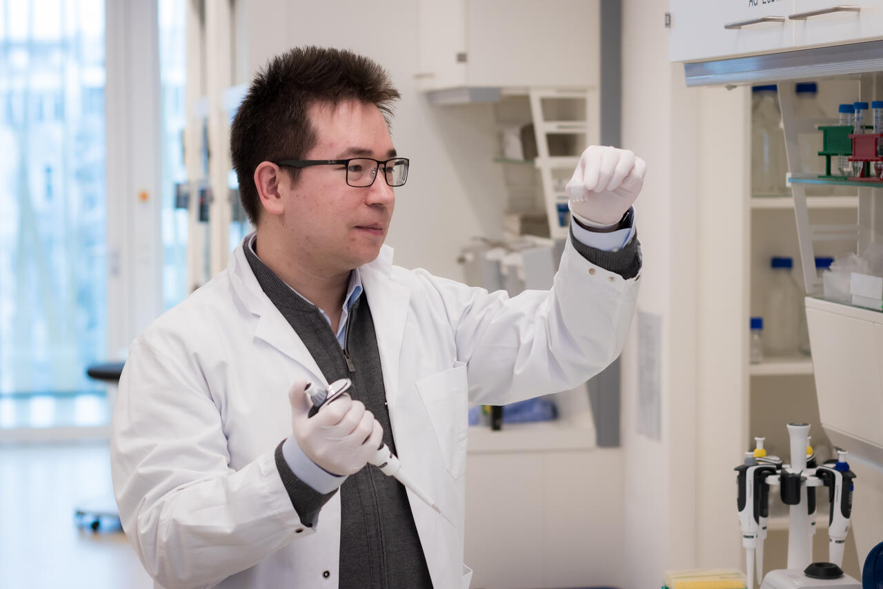
Leif Si-Hun Ludwig has been awarded a prestigious Heisenberg professorship in stem cell dynamics and mitochondrial genomics by the the Berlin Institute of Health at Charité, a position funded by the German Research Foundation (DFG).
Professor Leif Si-Hun Ludwig has led an Emmy Noether Research Group at the Berlin Institute of Health and the Max Delbrück Center since 2020. He has now been awarded a Heisenberg Professorship for Stem Cell Dynamics and Mitochondrial Genomics by the the Berlin Institute of Health (BIH) at Charité, a position funded by the German Research Foundation. Ludwig’s professorship will transition into a permanent role following an initial five-year funding period.
Studying how blood cells form
The human blood system is a marvel of constant regeneration, with millions of new cells replacing old blood and immune cells every second. These cells originate from hematopoietic (or blood-forming) stem cells in the bone marrow, maturing through several different developmental stages into the red and white blood cells, platelets, and B and T cells that keep us alive. While clinical blood tests can easily count these cells, determining the specific contribution of thousands of individual stem cells to overall blood production remains a significant challenge.
By observing natural mutations in human DNA, researchers can gain fundamental insights into how stem cells maintain healthy blood formation or how they behave when disease strikes. However, hunting for a single mutation within a genome of three billion base pairs is expensive, time-consuming and prone to error, even with today’s most advanced technology.
Cellular power plants reveal the origin of blood cells
To bypass these hurdles, Ludwig focuses on natural mutations within the mitochondrial genome – a much smaller, more manageable DNA molecule found within cellular power plants known as mitochondria. By pairing this approach with sophisticated single-cell sequencing technologies, his team can analyze tens of thousands of blood and bone marrow cells simultaneously, effectively mapping the activity of blood-forming stem cells. This single-cell analysis of natural genetic variation does more than track lineage; it provides vital information on the health of individual cells. In a clinical setting, this could eventually help doctors to predict the success of stem cell transplants or fine-tune cell and gene therapies with unprecedented precision.
Beyond this work on hematopoietic stem cells, Ludwig is tackling the challenge of inherited mitochondrial mutations. These defects are among the most common genetic disorders and can trigger a wide range of metabolic diseases affecting multiple organ systems. Despite their prevalence, the molecular causes of these conditions remain poorly understood. By investigating how mitochondrial gene variants influence different cellular and metabolic phenotypes, Ludwig aims to lay the groundwork for new therapeutic approaches.
“The Heisenberg program supports outstanding scientists, so it was no surprise to us that Ludwig was selected for this honor,” says Prof. Christopher Baum, Chair of the BIH Board of Directors and Chief Translational Research Officer of Charité. “His exceptional work combines basic research and application-oriented studies. Ludwig and his team are thus strengthening the translational network that brings together the BIH, Charité, and the Max Delbrück Center for the benefit of patients.”
Research / 06.02.2026
Professorship awarded to Mina Gouti
Mina Gouti has been awarded a professorship at Charité – Universitätsmedizin Berlin. The appointment will strengthen her pioneering organoid research at the Max Delbrück Center and deepen collaboration with clinicians to advance personalized medicine.
Dr. Mina Gouti, Group Leader of the Stem Cell Modeling of Development and Disease lab at the Max Delbrück Center, has been appointed W3 Professor of Complex Organoid Models for Personalized Medicine in the Medical Faculty, Charité – Universitätsmedizin Berlin. The permanent position recognizes her achievements in developing advanced organoid systems to understand how specific types of spinal cord neurons and skeletal muscle cells grow in space and time during development.
“This professorship will enable my team to fully leverage Berlin’s exceptional biomedical ecosystem and to work closely with clinicians at Charité to create complex organoids for personalized medicine,” Gouti says. This close interaction is essential for translating patient-derived organoid models into clinically relevant insights.”
The Gouti lab has developed three-dimensional neuromuscular organoids from human pluripotent stem cells that replicate key features of spinal cord neurons and skeletal muscle – providing powerful platforms for studying neuromuscular diseases such as spinal muscular atrophy and amyotrophic lateral sclerosis, and for drug screening. Such organoids can also be grown from cells derived from individual patients to model their specific disease.
The position reflects a shared vision between the Max Delbrück Center and Charité to accelerate translational research through fostering closer collaboration between clinicians researchers. “We now have the long-term perspective required to develop complex, functional organoids as predictive platforms for personalized medicine and early disease interception,” says Gouti. “We hope our research will lead to better health outcomes not only for people with neuromuscular diseases, but also those at high risk of developing them.”
Text: Gunjan Sinha
Further information
- News
- Events
- MAGAZINE „buchinside“
- Press service/Download
-
News Archiv
- Archive News 2026
- Archive News 2025
- Archive News 2024
- Archive News 2023
- Archive News 2022
- Archive News 2021
- Archive News 2020
- Archive News 2019
- Archive News 2018
- Archive News 2017
- Archive News 2016
- Archive News 2015
- Archive News 2014
- Archive News 2013
- Archive News 2012
- Archive News 2011
- Archive News 2010
- Archive News 2009
- Archive News 2008


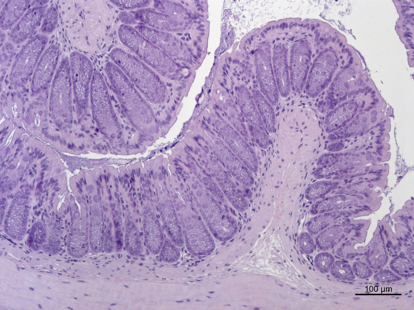